Download
1 / 38
390 likes | 482 Views
Explore the classification, pathogenesis, and treatment of Mycobacteria, including details on Tuberculosis and Leprosy. Key topics include epidemiology, laboratory diagnosis, and prevention strategies. Gain insights into this complex bacterium.

E N D
Genus = Mycobacteria By: Prof. A.M.Kambal Consultant Microbiologist & Head of the Bacteriology
Genus = Mycobacteria Definition: • Acid – Fast Bacilli / Acid and Alcohol fast bacilli, non – motile. Aerobic bacilli difficult to stain by gram stain - But stained by Z – N stain. Acid fastness due to Mycolic acid in cell wall.
Classification I- Parasitic a) M. tuberculosis complex: (i) M.t.b. (hominis) – humans (ii) M. bovis – cattle cause (iii) M. africanum – human tuberculosis (vi) Others b) M. leprae II- Saprophytic (Environmental) E.g.M. kansasii M. avium - intracellulare
M. tberculosis (M.t.b.) (hominis)(humans) • Optimum Temperature: 37 % • Takes 6-8 weeks on average to see colonies • Medium Complex. Commonly used one is egg – based called Loweinstein – Jensen (L.J). Pathogenesis: Causes TB mainly affect lung but also other organs. • Route – respiratory – inhalation of infected droplet nuclei.
Infection - Pulmonary TB 2 Types: I- Primary TB bacilli settle in the lungs usually the Right lung in the middle lobe. • Granulomatous inflammation • Lesion called tubercle in the sub pleural space • Spread to regional mediastinal lymph nodes • Similar lesions occur
Pulmonary TB (Continued) • Further spread of bacilli to other organs in the body • In 90 – 95 % of people with primary infection, the tubercle heals. • In the remaining 5-10 % the infection does not heal, secondary to immunosuppression and leads to active disease in multiple organs and is then referred to as miliary tuberculosis.
(II) Secondary TB (open / infectious) • Usually in the lungs especially apex of the right lung. It maybe by: a) Reactivation (Endogenous) - as a result of suppression of host”s defence system by: Diabetes Steroid and other immunosuppressive Malignant disease e.g. Leukemia
Secondary TB (Open Infections) Continued b) Reinfection (Exogenous) • In secondary TB. There is excessive tissue destruction of the lung leading to formation of cavities. Patient Complain of: a) Cough b) Loss of weight c) Fever d) Cough of blood, i.e. Haemoptysis • In secondary TB too, the disease process may become as progressive as in primary TB and lead to miliary TB.
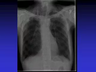
Tuberculosis: (a) Chest X-ray of a patient with tuberculosis bronchopneumonia. (b) Chest X-ray of the same patient 10 months after antituberculous therapy. (Courtesy of Dr. R.S.Kennedy)
Pathogenesis of Pulmonary Tb Infection Non-Immune Host (tuberculin –ve) Asymptomatic Primary Symptomatic Infection Primary Infection Tuberculin +ve Reactivation TB Resolution Progressive (Majority) Disease Apical Lung disease Bronchopneumonia Miliary TB TB Meningitis Urinary TB Bone TB Chronic Progressive Disease
Virulence factor • The bacilli survive and multiply in the macrophages Epidemiology:Pulmonary TB is a communicable disease. i) Sources of infection in the community are the open pulmonary lesions (usually seen in adults).
Epidemiolgy (Continued) (ii) Environmental Factors : - over crowding, poor ventilation and housing (iii) Populations at special risk: - Health workers – Doctors Nurses Laboratory Staff Immunosuppressed patients (iv)Underlying Diseases:Sarcoidosis Pneumoconiosis Diabetes
Laboratory Diagnosis i)Specimen - Sputum, Urine, CSF etc. 3 successive specimens ii)Direct Microscopy - Acid alcohol – fast bacilli by Z.N. stain of auramine fluorescent. Fluorescent Auramine Gel - More sensitive less specific iii) Culture - Loweinstein Jensen Media (4 – 8 weeks) iv)Guinea Pig - inoculation - Kill guinea pig after 6 – 8 weeks.
Treatment: • Prolonged, multiple and combined treatment schedule: i) Ethambutol (or Pyrazinamide) plus Isoniazid for 2 mos. plus Rifampicin ii) Then continue with Isoniazid and Rifampicin for 4 more months. iii) Total duration of treatment 9 months.
Prevention And Control Control: • Isolate all open pulmonary cases and treat effectively. • Trace all contacts of the index case by: a)Tuberculin testing: This is a T-cell mediated hypersensitivity reaction. Interpretation:Positive Tests: Patient has at least previously come into contact with M.tb and developed infection. Negative: Patient has not previously come into contact with a case of tuberculosis and has not therefore developed the primary infection.
Prevention • Vaccination: Live attenuated vaccine called Bacilli – Calmette Guerin (BCG) given to all newborn babies. N.B. B.C.G. not given to those already tuberculin positive. Immunity in T.B. is cell mediated and protection given by B.C.G. is about 10 years. 2)Chemoprophylaxis: Isoniazid is given to those who are tuberculin positive but who don’t have disease.
M. Leprae This is the cause of leprosy in humans. It affects the skin, peripheral nerves and the nasal mucosa. • M. leprae is weakly acid fast. Culture: Can not be grown in artificial media in the lab. Grown in animals: (i) Foot pad of mice or (ii) The armadillo
Infection is acquired mainly by inhalation of respiratory droplets. • Outcome of the infection depends on the status of patient’s cell mediated immune response: • Tuberculoid leprosy Seen in patients with competent T-cell function and there is slow and progressive development.
2)Lepromatous leprosy • In such patients the cell mediated immune system is very poor and the bacilli are therefore able to multiply and spread through blood without any inhibition by the patient’s immune system. The lesions are contagious. i.e. Infectious. • Disease is more severe in the Lepromatous form. Lepromin test:Similar to tuberculin test Diagnosis: Z/N stain of (i) Nasal scrapings or (ii) Ear lobe aspirate and look for AFB
Leprosy. Mutilation of fingers due to trophic changes associated with anaesthesia caused by infection of peripheral nerves.
Treatment: R (i) Dapsone - but many strains now resistant (ii) Dapsone + Rifampicin - Duration about 1-2 years. Vaccination: Combination killed M. leprae + B.C.G.
Epidemiology • Source of infection usually a case of leprosy • Requires close and prolonged contact for infection to occur • Believed that most infections are acquired in childhood
Laboratory Diagnosis i)Specimen - Sputum, Urine, CSF, etc. ii)Direct Microscopy - Acid Alcohol – fast bacilli by Z.N. stain of auramine & fluorescent. iii)Culture- Lowenstein Jensen Media (4-8 weeks) iv)Guinea Pig - Inoculation - Kill g. pig after 6-8 weeks.
Environmental Mycobacteria(Atypical / Saprophytic / Mycobacteria other than Tuberculosis (MOTT)) • Members are found in the environment E.g.Soil, water and others may be found in birds and other animals. • They all grow on L.J. media and some will also grow on Blood Agar. Some are slow growing just as M. tuberculosis but others grow fast within 5-7 days.
Environmental Mycobacteria (Continued) Some produce yellowish or orange pigment only when growing in the presence of light and are called PHOTOCHROMOGENS; others will produce pigment whether grown in light or darkness and are called SCOTOCHROMOGENS, and other do not produce any pigment at all. They are called NONCHROMOGENS. Atypical mycobacteria are opportunistic and do not produce disease in the normally healthy person.
Infections by these mycobacteria are not communicable 1) M. avium – intracellulare – cause tuberculosis in birds. Infections in human only under special circumstances in AIDS patients. It is slow growing Non chromogenic Causes pulmonary disease in immunocompromised specially AIDS patients. Often resistant to many anti TB drugs. 2) M. kansasii - usually found in soil. • Photochromogenic • Slow growing • Optimum temperature 37°C
Mycobacteria are not communicable (Continued) 3) M. scrofulaceum • Scotochromogenic • Slow growing • Optimum temperature 25°C 4) M. fortuitum • Fast grower – within 5-7 days • Causes infection in soft tissue. E.g. Skin, muscles, forming abscesses.
Negative Tuberculin Test • No induration, either due to: • no previous infection • pre-hypersensitivity stage • lost TB sensitivity with loss of Ag. • AIDS, anergic, susceptible to infection
Secondary TB (Continue) • Clinically: fever, cough, hemoptysis. • Source: - endogenous(reactivation) - exogenous (reinfection)
Immunity to Tuberculosis • Cell-mediated immunity associated with delayed hypersensitivity reaction. • Detected by tuberculin test. • Takes 2-10 weeks to react to tuberculin.
Negative Tuberculin Test • No induration, either due to: • no previous infection • pre-hypersensitivity stage • lost TB sensitivity with loss of Ag. • AIDS, anergic, susceptible to infection
Quantiferon G test • It measures the amount of interferon produced by lymphocytes in patients latently infected by M.tuberculosis • Compared to TST IT IS NOT AFFECTED by atypical mycobacterial infection