Download
1 / 35
390 likes | 703 Views
INFECTIONS OF THE GENITOURINARY TRACT. DEPARTMENT OF UROLOGY IAŞI – 20 13. INFECTIONS OF G-U TRACT. EPIDEMIOLOGY < 1 yr – bacteriuria: 2.7% M (phimosis), 0.7% F 1-5 yrs: 4.5% F, 0.5% M (congenital abnormalities; VUR or obstruction) 6-15 yrs (functional abnormalities: dysfunctional voiding)

E N D
INFECTIONS OF THE GENITOURINARY TRACT DEPARTMENT OF UROLOGY IAŞI – 2013
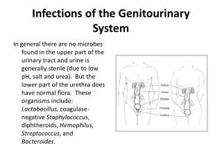
INFECTIONS OF G-U TRACT EPIDEMIOLOGY • < 1 yr – bacteriuria: 2.7% M (phimosis), 0.7% F • 1-5 yrs: 4.5% F, 0.5% M (congenital abnormalities; VUR or obstruction) • 6-15 yrs (functional abnormalities: dysfunctional voiding) • 16-35 yrs: F 20% (sexual intercourse and diaphragm use • 36-65 yrs: F (gynecologic surgery and bladder prolapse), M (prostatic hypertrophy/obstruction, catheterization and surgery) • > 65 years: (incontinence and chronic use of urinary catheters) PATHOGENESIS – bacterial entry (4) • ascending: periurethral bacteria urinary tract; short female urethra + close proximity to the vaginal vestibule and rectum
INFECTIONS OF G-U TRACT • hematogenous – in immunocompromised patients and neonates; Staph aureus, Candida sp and Mycobacterium tuberculosis • lymphatogenous – spread through the rectal, colonic, and periuterine lymphatics • direct extension – intraperitoneal abscesses, vesico-intestinal or vesico-vaginal fistulas; relapsing infection from an inadequately treated focus in the prostate or kidney Host Defenses • unobstructed urinary flow (washout of ascending bacteria) • urine specific characteristics (osmolality, urea concentration, organic acid concentration and pH) inhibit bacterial growth and colonization; factors that inhibit bacterial adherence (glycoprotein)
INFECTIONS OF G-U TRACT • presence of foreign bodies (stones, catheters, stents) allows the bacteria to hide from the host defenses • cells of the urinary tract secrete chemoattractants (interleukin-8) to recruit neutrophils to the area and limit tissue invasion • specific serum and urinary antibodies produced by the kidney bacterial opsonization and phagocytosis and bacterial adherence • normal flora of the periurethral area (lactobacillus) or the prostate (Zn) • in children, VUR allow bacteria to be inoculated into the upper tract and the infection to progress • aging: susceptibility – incidence of obstructive uropathy (M), alteration in the vaginal and periurethral flora (F), soiling of the perineum from fecal incontinence, neuromuscular diseases, increased instrumentation and bladder catheterization
INFECTIONS OF G-U TRACT Bacterial Pathogenic Factors • Escherichia coli – uropathogens = limited number of O, K and H serogroups increased adherence to uroepithelial cells [fimbriae (pili)], resistance to the bactericidal activity of human serum, production of hemolysin ( tissue invasion and makes iron available for the infecting pathogens) and expression of K capsular antigen (protects from phagocytosis by neutrophils) CAUSATIVE PATHOGENS • 80% of the uncomplicated cystitis and pyelonephritis –E coli; less common – Klebsiella, Proteus,Enterobacter spp and enterococci • hospital-acquired UTIs – a wider variety of causative organisms, including Pseudomonas and Staphylococcus spp • children – Klebsiella and Enterobacter spp
INFECTIONS OF G-U TRACT DIAGNOSIS • relies on urinalysis and urine culture, from a voided or bagged specimen, suprapubic aspiration or from a urinary catheter • occasionally, localization studies may be required to identify the source of the infection Urinalysis • rapid screen for UTIs (urine dipstick) – leukocyte esterase (white blood cells) and urinary nitrite • microscopic examination for WBCs (> 3 per HPF) and bacteria Urine Culture • quantitative culture for specific bacteria: > 100,000 CFU/mL (to exclude contamination) • clinically significant UTI can occur with < 100,000 CFU/mL bacteria
INFECTIONS OF G-U TRACT Localization Studies • upper urinary tract localization: bladder irrigated with sterile water, ureteral catheter placed into each ureter, specimen collected from the renal pelvis • in M, infection in the lower urinary tract (Meares and Stamey); specimen collected at the beginning of the void (urethra), midstream specimen (bladder), prostate massaged and void (prostate) ANTIBIOTICS • goal – to eradicate the infection by selecting the appropriate antibiotics that would target specific bacterial susceptibility
INFECTIONS OF G-U TRACT • general principles for selecting the appropriate antibiotics • infecting pathogen (antibiotic susceptibility, single-organism vs. poly-organism infection, pathogen vs. normal flora, community vs. hospital-acquired infection) • patient (allergies, underlying diseases, age, previous antibiotic therapy, other medications currently taken, outpatient vs. inpatient status, pregnancy) • the site of infection (kidney vs. bladder vs. prostate) • certain antimicrobial agents – adjusted in the presence of liver or renal diseases • in patients with recurrent UTIs or those at risk for UTI (children with VUR) – prophylactic antibiotics
INFECTIONS OF G-U TRACT • trimethoprim-sulfamethoxazole (TMP-SMX) – except Enterococcus and Pseudomonas spp; interferes with the bacterial metabolism of folate; highly effective and relatively inexpensive; adverse reactions: hypersensitivity reactions, rashes, gastrointestinal upset, leukopenia, thrombocytopenia and photosensitivity • fluoroquinolones – broad spectrum of activity, except Streptococci species and anaerobic bacteria; interfere with the bacterial DNA gyrase, preventing bacterial replication; highly effective but relative expensive; adverse reactions: mild gastrointestinal effects, dizziness and lightheadedness; should not be used in pregnant patients and in children (damage to developing cartilage)
INFECTIONS OF G-U TRACT • nitrofurantoin – good activity against most gram-negative bacteria (except Pseudomonas and Proteus spp), Staphylococci and Enterococci species; inhibits bacterial enzymes and DNA activity; highly effective and relative inexpensive; adverse reactions; gastrointestinal upset, peripheral polyneuropathy and hepatotoxicity, pulmonary hypersensitivity reaction and interstitial changes • aminoglycosides – used in the treatment of complicated UTI; highly effective against most gram-negative bacteria; combined with ampicillin, are effective against enterococci; inhibit bacterial DNA and RNA synthesis; adverse effects: nephrotoxicity and ototoxicity; regimen is directed toward obtaining higher peak and lower trough levels (more effective microbial killing while reducing toxicity)
INFECTIONS OF G-U TRACT • cephalosporins – good activity against most uropathogens; inhibit bacterial cell wall synthesis; adverse reactions: hypersensitivity and gastrointestinal upset; in children with febrile UTI/pyelonephritis, oral third-generation cephalosporins (cefixime) are safe and effective • aminopenicillins (amoxicillin and ampicillin) – good activity against Enterococci, Staphylococci, E coli and Proteus mirabilis; addition of ß-lactamase inhibitors (clavulanic acid) makes more active against the gram-negative bacteria; adverse reactions; hypersensitivity, gastrointestinal upset and diarrhea
INFECTIONS OF THE KIDNEY ACUTE PYELONEPHRITIS • inflammation of the kidney and renal pelvis, and its diagnosis is usually made clinically Presentation and Findings • chills, fever and costovertebral angle tenderness; often accompanying lower-tract symptoms (dysuria, frequency and urgency); sepsis may occur (20-30% of urosepsis) • E coli is the most common causative organism (80%), Klebsiella, Proteus, Enterobacter, Pseudomonas, Serratia and Citrobacter spp.; gram-positive bacteria (Streptococcus faecalis and S aureus) Imaging • renal US – rule out concurrent urinary tract obstruction; enlarged kidney, hypoechogenic parenchyma
INFECTIONS OF THE KIDNEY • CT scan (not necessary unless diagnosis is unclear or patient is not responding to therapy): constriction of peripheral arterioles and reduced perfusion of the affected renal segments (segmental, multifocal or diffuse – areas of reduced signal density), renal enlargement, attenuated parenchyma and a compressed collecting system • radionuclide study (99mTc-DMSA): detecting the perfusion defects of pyelonephritis Management • depends on the severity of the infection; toxicity because of associated septicemia hospitalization • empiric therapy – i.v. ampicillin and aminoglycosides or amoxicillin with clavulanic acid or a third-generation cephalosporin
INFECTIONS OF THE KIDNEY • parenteral therapy – until the patient defervesces; if bacteremia is present, parenteral therapy should be continued for an additional 7-10 days, then oral treatment for 10-14 days • patients who are not severely ill, outpatient treatment with oral antibiotics: fluoroquinolones or TMP-SMX for 10-14 days EMPHYSEMATOUS PYELONEPHRITIS • necrotizing infection – presence of gas within the renal parenchyma or perinephric tissue • 80-90% have diabetes; the rest – associated with urinary tract obstruction (calculi) or papillary necrosis Presentation and Findings • fever, flank pain and vomiting that fails initial management with parenteral antibiotics; pneumaturia may be present: bacteria – E coli, Klebsiella pneumoniae, Enterobacter cloacae
INFECTIONS OF THE KIDNEY Radiographic Imaging • diagnosis: gas overlying the affected kidney on a plain abdominal radiograph (KUB); CT scan – more sensitive in detecting the presence of gas in the renal parenchyma than renal US Management • essential – prompt relief of urinary obstruction (percutaneous drainage), control of blood glucose, fluid resuscitation and parenteral antibiotics • mortality: 11-54%; poor prognostic factors: high serum creatinine level, low platelet count, the presence of renal/perirenal fluid + bubbly/loculated gas pattern or gas in the collecting system • nephrectomy may be required; 3-4 weeks of parenteral antibiotic therapy is usually required
INFECTIONS OF THE KIDNEY RENAL/PERINEPHRIC ABSCESS • severe infection that leads to liquefaction of renal tissue, subsequently sequestered • rupture out into the perinephric space perinephric abscesses; extend beyond the Gerota's fascia paranephric abscesses • hematogenous spread of staphylococci (infected skin lesions) abscesses in the renal cortex • patients with diabetes, undergoing hemodialysis or i.v. drug abusers – high risk • abscesses due to gram-positive bacteria are less prevalent; those caused by E coli or Proteus species are becoming more common – formed in the corticomedullary junction, in conjunction with underlying urinary tract abnormalities (stones or obstruction)
INFECTIONS OF THE KIDNEY Presentation and Findings • fever, flank or abdominal pain, chills and dysuria • flank mass may be palpated • urinalysis – usually WBCs; normal in approx. 25% of the cases • urine cultures – 1/3; blood cultures – 1/2 Imaging • US – anechoic mass within or displacing the kidney/echogenic fluid collection that blends with the fat within Gerota's fascia • CT scan– enlarged kidney with focal areas of hypoattenuation mass with a rim of contrast enhancement ("ring" sign); thickening of Gerota's fascia, stranding of the perinephric fat or obliteration of the surrounding soft-tissue planes
INFECTIONS OF THE KIDNEY Management • appropriate antibiotic therapy– empiric therapy with broad-spectrum antibiotics (ampicillin or vancomycin + aminoglycoside or third-generation cephalosporin) • w/o respose within 48 h percutaneous drainage under CT or US guidance culture of the drained fluid • still not resolved open surgical drainage or nephrectomy • evaluation for underlying urinary tract abnormalities (stone or obstruction) XANTHOGRANULOMATOUS PYELONEPHRITIS • form of chronic bacterial infection of the kidney – hydronephrotic and obstructed severe inflammation and necrosis of the kidney parenchyma
INFECTIONS OF THE KIDNEY • foamy lipid-laden histiocytes (xanthoma cells) renal clear cell carcinoma Presentation and Findings • history of urolithiasis (35%) • flank pain, fever, chills and persistent bacteriuria • physical examination – flank mass often palpated • urinalysis – WBCs and protein, urine culture – E coli, Proteus • anemia, hepatic dysfunction (50%) Imaging • CT scan (most reliable) - large heterogeneous, reniform mass; renal parenchyma marked with multiple water-density lesions (dilated calyces or abscesses); inflammatory process extend to perinephric fat, retroperitoneum and adjacent organs (psoas muscle, spleen, colon or great vessels)
INFECTIONS OF THE KIDNEY • renal US – enlarged kidney with a large central echogenic area and anechoic parenchyma • misdiagnosed as a renal tumor – similar appearances Management • nephrectomy diagnosis is made pathologically PYONEPHROSIS • bacterial infection of a hydronephrotic & obstructed kidney suppurative destruction of renal parenchyma (loss of renal function) • sepsis may rapidly ensue rapid diagnosis and management Findings • high fever, chills, flank pain& pyuria
INFECTIONS OF THE KIDNEY • bacteriuria & leukocyturia(may be absent with complete obstruction!) • US – persistent echoes in the lower part of the collecting system, fluid-debris level with echoes that shift with positional changes, strong echoes with acoustic shadowing (air in the collecting system), dilated collecting system, renal or ureteral calculi • IVU – opacities, nonfunctional kidney Management • immediate institution of antibiotic therapy and drainage of the infected collecting system (percutaneous nephrostomyor ureteral stent) • then, treatment of the cause (urolithiasis, UPJ obstructionetc.) or nephrectomy
INFECTIONS – BLADDER ACUTE CYSTITIS • urinary infection of the lower urinary tract (bladder); F > M • irritative voiding symptoms (dysuria, frequency & urgency) • low back and suprapubic pain, hematuria, and cloudy/foul-smelling urine • urinalysis – WBCs, hematuria; urine culture • management – short course of oral antibiotics (TMP-SMX, nitrofurantoin, fluoroquinolones) – 3-5 d RECURRENT CYSTITIS/UTI • caused by bacterial persistence ( removal of the infected source) or reinfection with another organism ( preventive therapy)
INFECTIONS – BLADDER • bacterial persistence imaging (US, IVU, cystoscopy, CT scan, bacterial localization studies, retrograde pyelograms) • bacterial reinfection evidence of vesicovaginal or vesicoenteric fistula Management • bacterial persistence surgical removal of the infected source (urinary calculi) • bacterial reinfection prophylactic antibiotics (low-dose continuous or intermittent self-start), surgical repair of fistulas • related to sexual activity frequent emptying of the bladder & single dose of antibiotic, after intercourse • intravaginal estriol, lactobacillus vaginal suppositories and cranberry juice taken orally
INFECTIONS – PROSTATE ACUTE BACTERIAL PROSTATITIS • inflammation of the prostate associated with a UTI ascending urethral infection or reflux of infected urine from the bladder into the prostatic ducts Presentation and Findings • abrupt onset of fever, chills, malaise, arthralgia, myalgia, lower back/rectal/perineal pain and urinary symptoms (frequency, urgency, dysuria acute urinary retention) • DRE – tender, enlarged irregular and warmgland • urinalysis – WBCs, occasionally hematuria • leukocytosis; PSA • ! urethral catheterization & prostatic massageshould be avoided bacteremia
INFECTIONS – PROSTATE • US – residual urine; TRUS – non-responsive to conventional therapy Management • trimethoprim or fluoroquinolones (high drug penetration into prostatic tissue) for 4-6 wks. (prevent complications – chronic prostatitis, abscess formation) • sepsis, immunocompromised pts., acute urinary retention or significant medical comorbidities hospitalization and parenteral antibiotics(amoxyclav + aminoglycoside) • urinary retention suprapubic catheter CHRONIC BACTERIAL PROSTATITIS • relapsing, recurrent UTI caused by the persistence of pathogen in the prostatic fluid, despite antibiotic therapy
INFECTIONS – PROSTATE • dysuria, urgency, frequency, nocturia and low back/perineal pain • others are asymptomatic, but have bacteriuria • DRE is often normal; occasionally, tenderness, firmness or prostatic calculi • urinalysis – WBCs and bacteriuria; PSA may be • diagnosis – identification of bacteria from prostate expressate or urine specimen after a prostatic massage (4-cup test) • TRUS – if prostatic abscess is suspected Management • antibiotic therapy – similar to acute bacterial prostatitis, but up to 3-4 mo. • alpha blocker – to reduce symptom recurrences
INFECTIONS – PROSTATE • cure is not often achieved poor penetration of antibiotic into prostatic tissue & isolation of the bacterial foci within the prostate • recurrent episodes of infection suppressive antibiotic (TMP-SMX 80/240 mg daily, nitrofurantoin 100 mg daily, or ciprofloxacin 250 mg daily) • refractory disease ? TUR-P EPIDIDYMITIS • most cases < 35 years – due to sexually transmitted organisms (N gonorrhoeae, C trachomatis); in children and older men – E coli • epididymis testis
INFECTIONS – PROSTATE Presentation and Findings • severe scrotal pain – may radiate to the groin or flank; scrotal enlargement (inflammation of epididymis/testis or reactive hydrocele); symptoms of urethritis, cystitis or prostatitis • physical examination – enlarged and red scrotum; thickened spermatic cord • urinalysis – WBCs and bacteria in the urine or urethral discharge; blood analysis – leukocytosis • epididymitis acute testicular torsion • scrotal Doppler US – presence of blood flow in the testis • radionuclide scanning– uptake of the tracers into the center of the testis
INFECTIONS – PROSTATE • scrotal US – enlarged epididymis with increased blood flow; reactive hydrocele or testicular involvement Management • antibiotic treatment • gonococcal ceftriaxone (250 mg i.m.) or fluoroquinolones (ciprofloxacin 250 mg or norfloxacin 800 mg) • nongonococcal tetracycline or erythromycin (500 mg 4 times daily) or doxycycline (100 mg twice daily) for 7-14 days • bed rest, scrotal elevation, nonsteroidal anti-inflammatory agents • treatment of the sexual partner • abscess open drainage • chronic, relapsing epididymitis, scrotal pain epididymectomy
SPECIFIC INFECTIONS • specific infections – caused by specific organisms, clinically unique disease, specific pathologic tissue reactions TUBERCULOSIS • young adults (60% of pts. – age 20-40); M > F Etiopathogenesis • Mycobacterium tuberculosis • lungs hematogenous route GU organs • kidney bladder • prostate bladder,epididymis testis • renal parenchyma (no symptoms) calyces pus and organisms discharged into urine symptoms (of cystitis)
SPECIFIC INFECTIONS • infection of the pelvic mucosa and the ureter stricture and (uretero)hydronephrosis • caseous breakdown of renal tissue + Ca laid down in the reparative process • fibrosis of ureter shortened and straightened "golf-hole" ureteral orifice (incompetent valve) • bladder – vesical irritability; tubercles form, coalesce & ulcerate (bleeding); fibrosis & contraction of the bladder (marked frequency); ureteral reflux or stenosis ureterohydronephrosis • extensive epididymal infection abscess formation spontaneous rupture permanent sinus of the scrotal skin
SPECIFIC INFECTIONS Pathology • granuloma (basic lesion in TB) – aggregation of histiocytic cells(vesicular nucleus and clear cellbody), that can fuse with neigh-boring cells epithelioid reticulum;at the periphery are large cells withmultiple nuclei (giant cells) • virulence of organism resistanceof patient caseation and cavitation healing by fibrosis and calcification • bladder – tubercles can be seen endoscopically (white or yellow raised nodules surrounded by a halo of hyperemia)
SPECIFIC INFECTIONS Clinical findings • symptoms – vesical in origin (cystitis) • nonspecific complaints – generalized malaise, fatigability, low-grade persistent fever, night sweats • epididymis – painless or mildly painful swelling (including vas deferens), chronic draining sinus • evidence of extraGU tuberculosis (lungs, bone, lymph nodes, tonsils, intestines) Laboratory • persistent pyuria, acid pH, without organisms on usual cultures • acid-fast stain (Ziehl-Neelsen), cultures (Löwenstein-Jensen)
SPECIFIC INFECTIONS X-Ray findings • KUB – calcifications in the renal parenchyma • IVU – “moth-eaten” ulcerated calyces; obliteration of calyces; (U)HN due to ureteral stenosis from fibrosis; abscess cavities that connect with calyces; multiple ureteral strictures, with shortening and straightening of the ureter; non-functional kidney due to complete ureteral occlusion or renal destruction (autonephrecto-my) retrograde ureteropyelogram • US, CT Instrumental examination • cystoscopy – tubercles or ulcers biopsies + pathology
SPECIFIC INFECTIONS Treatment • Medical (2-3 m, 7/7 + 4-3 m, 2-3/7) • isoniazid (INH), 200–300 mg orally once daily; • rifampin (RMP), 600 mg orally once daily; • ethambutol (EMB), 25 mg/kg daily for 2 months, then 15 mg/kg orally once daily; • streptomycin, 1 g intramuscularly once daily; • pyrazinamide, l.5–2 g orally once daily. • Surgical – urinary diversion or augmentation cystoplasty (ileocystoplasty, ileocecocystoplasty, sigmoidocystoplasty), nephrectomy, epididymectomy