Download
1 / 78
1.41k likes | 3.64k Views
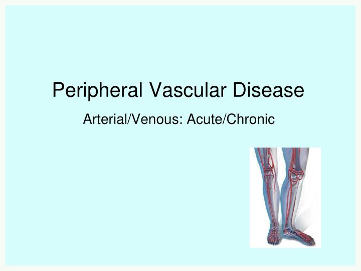
Peripheral Vascular Disease. Arterial/Venous: Acute/Chronic. Peripheral Vascular Disease. A term used to describe a group of diseases that involve pathophysiological changes in the “peripheral” arteries (i.e., excluding the coronary arteries) or veins resulting in blood flow disturbances.

E N D
Peripheral Vascular Disease Arterial/Venous: Acute/Chronic
Peripheral Vascular Disease • A term used to describe a group of diseases that involve pathophysiological changes in the “peripheral”arteries (i.e., excluding the coronary arteries) or veins resulting in blood flow disturbances.
Lymphatic disorders (also considered a form of PVD) Lymphatic system (ducts and nodes) • plays a role in filtering foreign particles • provides the only means by which interstitial proteins can return to the venous system (if blocked, obstructed [or *removed], edema may develop with risk of infection)
Raynaud’s Disease • characterized by bilateral intermittent arteriolar vasoconstriction/vasospasm in the hands and feet, often precipitated by emotional factors, cold, tobacco, causing color (white to blue to red) and temperature changes as well as burning pain in affected digits • is usually associated with underlying systemic disease (e.g., autoimmune disorders) • unlike other acute arterial disorders Raynaud’s is, with proper management (e.g., avoidance of triggers), essentially benign and self-limiting
Management • Thought to be vasospasm resulting from an exaggerated response to SNS stimulation • calcium channel blockers (i.e. nifedipine (Adalat), diltiazem *Cardizem) may be used) • Prevention • avoid/manage stress • avoid exposure to cold/cold H20 • avoid nicotine, caffeine, drugs that elicit a vasoconstrictive effect • safety precautions
Acute Arterial Insufficiency(Acute Arterial Occlusion) • usually involves complete blockage, is of sudden onset, and constitutes an emergency situation (muscle necrosis within 2-3 hours) • etiologies include: • arterial compression • thrombosis/embolism • arterial injury/damage • Risk factors • nonmodifiable; modifiable Risk factors: Age Gender (male) Family history Modifiable risk factors: Hypertension Diabetes Hyperlipidemia Smoking Obesity
Thrombosis • The formation of a blood clot within a blood vessel • Can occur in the arterial or venous systems • Leads to obstruction of a blood vessel in the circulatory system • Can lead to ischemia and infarction, and even death • Can also lead to embolism • Clot within a vessel breaks free and travels through body (“embolizes”) • Thromboembolism is combination of a thrombosis and embolus
Why does this happen? • Hemostasis • Formation of blood clot formation at the site of vessel injury • Carefully regulated system • Involves platelets and coagulation factors • Lack of coagulation factors bleeding • Overactive coagulation cascade thrombosis
Arterial thrombosis (ie. ischemic limb)
Fixed mottling & cyanosis What are the features of an acute ischemic limb? REMEMBER THE 6 P’S: • PAIN • PALLOR • PULSELESNESS • PERISHING COLD (POIKILOTHERMIA) • PARASTHESIAS • PARALYSIS excruciating pain color pale or cyanotic may be pulseless skin cool to touch loss of sensation/ position sense loss of movement
Chronic Arterial Insufficiency(Peripheral Arterial Disease) • primarily caused by atherosclerosis,disrupting the balance between arterial oxygen supply and demand • risk factors same as for CAD, with diabetes, HTN, and smoking as particularly high risk factors(see Table 40-3 Collaborative Care: Peripheral Arterial Disease
Peripheral Arterial Disease • Thickening of artery walls • Progressive narrowing of the arteries of the upper and lower extremities • resulting in tissue perfusion and ischemia in the area distal to the obstruction • may be acute or chronic
Risk Factors • Typical Patient: • Smoker (2.5-3x) • Diabetic (3-4x) • Hypertension • Hx of Hypercholesterolemia/AF/IHD/CVA
Patients at risk — Based in part upon the above observations, the 2005 American College of Cardiology/American Heart Association (ACC/AHA) guidelines on PAD, which were produced in collaboration with major vascular medicine, vascular surgery, and interventional radiology societies, identified the following groups at risk for lower extremity PAD • Risk Factors: • Atherosclerosis (same as RF’s for CAD and CVD) • Smoking (2.5-3x) • Diabetes 3-4x • Hypertension, increased age >50, male and family history • RARE: homocysteinuria
Peripheral Arterial Disease • one limb is usually affected more than the other, therefore always compare bilaterally • lower limbs more susceptible than the upper limbs • most common locations for stenosis are the aortoiliac bifurcation and the femoral bifurcation • nearly half of the clients with arterial PVD have associated CAD
Clinical Manifestations • intermittent claudication (pain in calf, thigh, or buttock depending on the location of the blockage) • rest pain in forefoot especially at night (advanced disease) • capillary refill • elevational pallor • dependent rubor • “pins & needles” sensation (paresthesia, due to ischemia) • affected limb cool to touch
Peripheral Arterial Disease • weakened or diminished pulses • chronic trophic changes • thin, shiny, dry or scaling skin with hair loss • thick, yellow, & brittle toenails • muscle atrophy • ulceration (*arterial), gangrene possible • bruit • Erectile dysfunction (e.g., aortoiliac disease) • *please note the differing characteristics of arterial and venous ulceration in course text (Table 40-2)
ULCER • associated with claudication + signs of ischaemia • occur on dorsum of foot + anterior skin • ↓ pulses, cold to touch, hairless skin • Painful, punched out edge
Diagnosis • history (symptoms & presence of risk factors) • physical exam (signs) • doppler assessment of peripheral pulses • ankle/brachial index [ABI] (normal is ~1) • see p. 1015 • treadmill (exercise testing) • angiography (necessary before any surgery)
ABI Clinical Correlation • >0.9 • Normal Limb • 0.5-0.9 • Intermittent Claudication • <0.4 • Rest Pain • <0.15 • Gangrene (BP higher in ankle than arm)
Take the highest measurement in both limbs • low ABI is also predictive of an increased risk of all-cause and cardiovascular mortality and of the development of coronary artery calcification • 95% sensitive in detecting angiogram positive disease and around 99% specific in identifying supposedly healthy subjects
Goals of management • reduce progression of the disease • promote arterial blood flow • promote vasodilation • prevent vascular compression • pain relief • attain/maintain tissue integrity • promote adherence
Management • risk reduction/reduce disease progression • smoking cessation • weight reduction • exercise (e.g., walking), unless contraindicated • reduction of blood lipid levels • diet (e.g., cholesterol, saturated fats and TFAs, fiber) and, in some cases, pharmacologic intervention (e.g., antilipidemeic drugs) • blood glucose and blood pressure control • antiplatelet meds
promote arterial blood flow • keep lower extremities below the level of the heart (e.g., reverse trendelenburg position) • encourage, or assist with, walking or graded isometric exercise to increase collateral circulation (*if not contraindicated) • avoid prolonged standing or sitting in one position • pharmacologic therapy • Inhibit platelet aggregation
promote vasodilation and prevent vascular compression • apply external warmth (e.g., socks, warm bath, or warm drink), promoting tissue perfusion • NEVER APPLY DIRECT HEAT AS IT MAY CAUSE A BURN • prevent exposure to cold and chilling • avoid crossing legs/constrictive clothing and accessories • smoking cessation • minimize stressful situations
pain relief • analgesics (opioids) • maintain tissue integrity • prevent infection/injury/trauma • meticulous foot care • well-balanced diet that includes adequate protein • attain and maintain ideal weight • Promote self care • patient/client education
Surgical/Radiological Management • Management depends on the etiology and may include: • embolectomy • revascularization • anticoagulation • fibrinolytic agents • amputation • Prevention best • know those at risk and monitor them closely
percutaneous transluminal angioplasty (PTA) with stent insertion (e.g., isolated lesion) • endarterectomy (e.g., carotid artery) • thrombolytic therapy (acute emboli/arterial graft occlusion) • arterial bypass (vascular) grafting (e.g., femoral-popliteal graft using saphenous vein) • followed by anticoagulation/antiplatelets • amputation (in presence of gangrene)
Femoropopliteal Bypass (Fem-Pop Bypass) for Peripheral Arterial Disease
Venous Disorders • Acute Venous Disorders • superficial thrombophlebitis • thrombophlebitis/deep vein thrombosis (DVT)/PE • Chronic Venous Disorders • varicose veins • chronic venous insufficiency
Venous thromboembolism Deep vein thrombosis Pulmonary embolism
Venous thromboembolism • Deep venous thrombosis • Blood clot in the proximal veins of the leg • Less commonly in the arms • Symptoms include: • Pain (never massage) • Swelling (calf circumference) • Redness • Warmth • PE could be first manifestation! • Above affecting one limb (unilateral)! Most common in lower extremities
most common in the lower extremities • 50% may be asymptomatic • unilateral swelling distal to the site (elevated venous pressure from venous pooling pushes fluid into interstitial spaces creating edema) *[may need to measure circumference] • *pain on dorsiflexion (Homan’s sign) is present in less than 1/3 and is no longer considered a valid sign for DVT • tenderness to palpation of calf (never massage!) • redness or warmth of the leg • dilated (prominent) veins • low-grade fever • unfortunately, pulmonary embolism may be the first clinical manifestation for some
Venous thromboembolism • Pulmonary embolism • Blood clot (from DVT) breaks off • Travels to lung • Can lead to infarct • Symptoms: • Chest pain • Shortness of breath • Lightheadedness (low BP) • Syncope • Hemoptysis • Can be life threatening!
Pulmonary embolism • Untreated PE • Mortality rate of ~30%1 • Most die within hours of diagnosis • Treated PE • Prospective NEJM study looked at 399 patients with newly diagnosed PE • 94% received conventional treatment • Only 2.5% (10 patients) died of PE • Treatment of PE is life-saving!
Venous thromboembolism • Incidence estimated at 1-2 in 1000 • Known predisposing conditions – Virchow’s triad: Venous stasis Alterations affect the balance between bleeding and clotting Vessel wall injury Hypercoagulability
Venous stasis: immobility or absence of calf muscle pump, paralysis, stroke, anesthesia, immobility due to bedrest, prolonged travel, obesity, pregnancy, restrictive clothing, reduced blood flow, CHF, shock, vasodilation • Hypercoagulability: abrupt withdrawal from anticoagulants, accompanies some malignant neoplasms (especially visceral and ovarian tumors), dehydration, blood dyscrasias (e.g., polycythemia vera), sepsis, use of oral contraceptives (especially in combination with smoking), HRT • Vessel wall injury: physical/traumatic or chemical irritation to the vein (e.g., IV insertion/ IV medications & solutions), fractures and dislocations (e.g., hip), severe blows to an area, diseases of the vein, abdominal, pelvic, hip, or knee surgery
Venous thromboembolism • Risk factors: • Recent surgery (OR:25 (10-50) • Immobility • Trauma • Hormones (OCP’s, HRT) • Pregnancy • Previous DVT/PE • Family history • Cancer • Be aware of a “perfect storm” Thromboprophylaxis is effective, and every inpatient should be risk-stratified….this includes nurses asking the questions!!!!
previous DVT (must identify high risk situations and initiate prophylaxis, for example, LMWH) • thrombus formation is usually attributed to two or more factors of Virchow’s triad: venous stasis hypercoagulability injury to the venous wall
Superficial Thrombophlebitis • clot formation obstructing venous flow in the superficial veins • may be iatrogenic: IV catheters or the instillation of caustic chemicals (potent drugs like antibiotics), contrast media, TPN • best to prevent (e.g., central line usage, revisit the standard nursing care for a client receiving IV therapy!) • What do you do? Iatrogenic: caused by medical treatment Remove IV, elevate hand, warm, moist heat, NSAIDs, sometimes anticoagulants
Medical Management • anticoagulation (prevent clot formation or clot extension) • unfractionated heparin IV (continuous drip or intermittent infusion) or SC • low-molecular weight heparin (LMWH) • warfarin [Coumadin] • thrombolytic therapy
Treatment of DVT/PE • Goals of treatment: • Short term: • Prevent the extension of thrombus and embolization of DVT • Reduce mortality for PE by reducing recurrent events • Relief of symptoms • Long term: • Prevent recurrent events
Treatment of VTE General Principles • Parenteral heparin for minimum 5 days • Initiate oral anticoagulants (ie. warfarin) on day 1 • May stop heparin once INR >2.0 for 24 to 48h • Consider outpatient therapy • No comorbidities requiring hospitalization • No hypoxia or chest pain • Need careful follow-up
Heparin vs LMWH Heparin (unfractionated) LMWH SQ, once daily or BID No need for routine monitoring (predictable pharmacokinetics) More expensive Useful Outpatient therapy Poor venous access • Needs IV admin • Needs aPTT testing using nomogram • Inexpensive • Useful • High risk for bleeding or requiring surgery • Renal failure • Antidote (protamine sulfate)


![Diffuse Vascular Disease (Focus on Peripheral Arterial Disease [PAD])](https://cdn1.slideserve.com/3363465/slide1-dt.jpg)