Download
1 / 9
90 likes | 113 Views
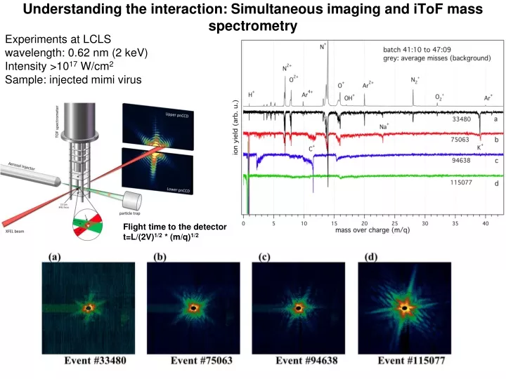
Experiments at LCLS wavelength: 0.62 nm (2 keV) Intensity >10 17 W/cm 2 Sample: injected mimi virus. Understanding the interaction: Simultaneous imaging and iToF mass spectrometry. Flight time to the detector t=L/(2V) 1/2 * (m/q) 1/2.

E N D
Experiments at LCLS wavelength: 0.62 nm (2 keV) Intensity >1017 W/cm2 Sample: injected mimi virus Understanding the interaction: Simultaneous imaging and iToF mass spectrometry Flight time to the detector t=L/(2V)1/2 * (m/q)1/2
Correlate diffraction to dynamics in a single measurement Incident pulse Plasma simulations: temperature, ionization Proton acceleration H+ Virus model (shape, composition) Hit strength Mass over charge (m/q) Scattered photons Proton acceleration Heating & explosion Proton flight time Preliminary results show amazing agreement
H+ Nb5+ Solid Nb sample C/O HP filter at 1 keV Nb4+ Nb3+ Nb2+ Nb1+ High field experiments: ToF measurements in drift mode Experiments at FLASH wavelength: 13.5 nm Intensity >1017 W/cm2 Sample: Solid niobium - Saturation in maximum observed proton energies - Generated by a transient surface transparency A grid in front of the detector can be charged and works as a high-pass filter. Physical Review E 83, 016403 (2011)
Craters in Deuterated vanadium studied using SEM and AFM Crater depth compared with maximum observed proton energies ss29, z-3.8 28 31 28’ 34,35 29,30 32,33 27’ ss28, z-3.6 25’ 26 25 26’ 27 21 ss27, z-3.4 22 23 24 20 18’ ss26, z-3.3 19’ 19 18 17 ss25, z-3.0 11 12 13 14 15 16 ss24, z-2.7 03 10 07 04 08,09 05,06 Experiments and analysis done together with Libor Juha et al. In situ focus determination using the maximum detectable proton energy. Resolution better than the Rayleigh length of the focusing mirror High Energy Density Physics, vol. 7, No. 1, 336-342 (2011)
Single atoms / molecules 20 fs pulses Ne charge state 1 2 8 9 10 200 fs pulses 200 fs pulses (neon atoms) -Hollow molecules at the highest pulse intensity and shortest pulse durations. -Decreases the risk of damage in single pulse imaging experiments since the damage depends on photo-ionization but the elastic scattering cross-section does not
FUNDAMENTAL ELECTRONIC PROCESSES at low and high photon energies Photo e- X-RAY PHOTON CARBON C2+ relaxed ion C+ hollow ion 2pz Auger e- t1/2 = 11.2 fs 2py 2s 1s 1s 2px INNER SHELL 1s 2s 2px 2py 2pz 1s 2s 2px 2py 2pz 1s 2s 2px 2py 2pz UV PHOTON CARBON C+ ion 2pz Photo e- 2py 2s 1s 2px OUTER SHELL 1s 2s 2px 2py 2pz 1s 2s 2px 2py 2pz
Molecular alignment: LCLS 2009 Time-Resolved Spectroscopy at the LCLS, Optics Express, Vol. 18, No. 17, 17620 (2010) J. M. Glownia, J. Cryan , J. Andreasson, et al. N22+(v=0) 50 fs N22+(v=0) N+(v) N+(-v) N22+(v=0) The alignment is revealed in the m/z region around 14 where the central N22+ peak is flanked by signal from N+ ions with initial velocity towards (v) and away from (-v) the detector. Processed data shows dynamics on 50 fs time scale Ratio: side peak / central peak • 800 nm Ti:Sapphire laser is used to impulsively align molecular nitrogen along the laser polarization direction • 1080 eV x-rays then ionize the N2 producing prompt coulomb explosion X-ray delay
Outlook: ToF instrumentation -VMI and sample orientation for 3D reconstruction - MD simulations show that on protein fragmentation is reproducible - Use ToF measurements with momentum resolution to get sample orientation and aid 3D image reconstruction Sulphur ions from exploding lysozyme proteins follow reproducible trajectories Top: schematic view of a VMI instrument. Bottom: Oxygen ions with four different initial velocities forming four rings on the detector in an experiment on ozone (images from Wikipedia) Reminder: there is also electronToF spectroscopy
Summary, sample Injector and Diagnostic Systems - What could we build and develop ? • Aerodynamic lens sample injector and catcher system • Laser-based injector and sample alignment system • Sample or pulse on demand systems • Fluorescence probe to characterize sample integrity during injection • Optical probes to record plasma emission at the interaction point • Ion-TOF instrumentation to characterize X-ray focus and sample fragmentation • Velocity Map Imaging (VMI) for measuring sample orientation from fragmentation • Electron-TOF instrument for measuring ionization dynamics (designed to be used singly or in coincidence with the ion-TOF or VMI) • Data acquisition system for flight time measurements • Native mass spectrometer (interfaced with the particle catcher/injector) for high-mass measurements Thank You