Download
1 / 36
410 likes | 560 Views
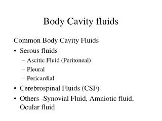
Body Fluids. Dr. Sangeetha Balakrishnan 23 February, 2017. Presumptive/Preliminary/Screening/ Field/Indicative Tests for Blood. Luminol Test Fluorescein Test Phenolpthalein Test. Luminol Test at a Crime Scene. 0 to 30 s. Fluorescein Test at a Crime Scene. Phenolpthalein Test.

E N D
Body Fluids Dr. Sangeetha Balakrishnan 23 February, 2017
Presumptive/Preliminary/Screening/Field/Indicative Tests for Blood • Luminol Test • Fluorescein Test • Phenolpthalein Test
Luminol Test at a Crime Scene 0 to 30 s
Phenolpthalein Test • Can detect blood diluted to 1 part in 10 million. • Gives false positives for certain vegetable extracts. • How is it done?: (i) Suspected blood stain (on a cotton swab) + water + Phenolpthalein colourless (ii) Add hydrogen peroxide immediate PINK colour!
Confirmatory Tests for Blood 1. Teichmann Test 2. Takayama Test (Hemochromogen Test) • Both are called Microcrystal Tests. Blood + Crystallising Reagent Characteristic Shaped Crystals.
Teichmann Test • Blood + Teichmann’s Reagent (heat) Hb hemin react with halides brownish-yellow rhomboid crystals • Microscopic Observation • Teichmann’s Reagent: Potassium bromide + Potassium chloride + Potassium iodide in Acetic acid.
Takayama Test (Hemochromogen Test) • Suspected blood stain on a glass slide + heat + add pyridine in Sodium hydroxide + reducing sugar red feathery crystals of pyridine ferroprotoporphyrin • Very sensitive test. • Even very old blood stains give +’ve results!
Saliva • Saliva is produced in the mouth. • Function: preliminary digestion of food. • Contains: water, proteins, enzymes, salts. • Alpha amylase: enzyme in saliva. • Alpha amylase breaks down starch in food. • It is also present in other body fluids, but in low concentrations.
Preliminary Test - Saliva • Principle: Starch + Iodine Deep blue colour • Suspected saliva sample + water/saline incubate at body temperature. • If the suspected sample is indeed saliva, it will contain alpha amylase! • Alpha amylase will break down starch into simpler components. (digestion) • Add Iodine solution. • Absence of starch (it has been broken down) No Deep Blue colour!
Drawbacks of the Saliva Preliminary Test • Not particularly sensitive. • Not specific to saliva. • Use saliva sample for DNA testing. • Detection of saliva at scene of crime: Shine UV light fluorescence!
Saliva – Confirmatory test • Phadebas Amylase Test Developed by Pharmacia Diagnostics. Qualitative and quantitative test. Phadebas: a synthetic biochemical substrate. • The substrate has starch microspheres. • The microspheres are chemically bonded to a blue coloured dye. Phadebas substrate + suspected saliva (in water) salivary amylase digests starch starch microspheres break down blue dye is released!
Urine • Main components: (i) Creatinine (ii) Urea Presumptive Test – Urine 1. Jaffe Test • Based on the detection of creatinine • Suspected urine sample + picric acid + 5% NaOH immediate orange colour
Presumptive Test – Urine … cont’d 2) Assay for Urea • Bromothymol Blue is a pH-indicator dye. • Colour: Yellow-green at pH 6 Aqua blue at pH > 7.6 • Whatman Filter Paper + Bromothymol Blue (1 drop) allow to dry • Add a drop from the suspected urine sample. • Add a drop of Urease. • If urea is present (meaning: urine is present), urease will degrade urea to ammonia gas. • This will cause the pH to increase. • Aqua blue colour!
Semen • Produced by the male sex organ. • Semen = Spermatozoa + Seminal fluid (cellular component) (fluid component)
Presumptive Test - Semen • Based on Seminal Acid Phosphatase (SAP). • There are other acid phosphatases in the body! • Brentamine Spot Test or Walker Test • Suspected Semen Sample + Brentamine Fast Blue B Intense purple colour within 2 minutes
Confirmatory Test - Semen • Christmas Tree Stain Test • Basis: Sperms analysed in lab are NOT motile. • Hence identification should be in the presence of other cellular material! • A pair of dyes is used: • Picroindigocarmine (PIC) • Nuclear Fast Red • PIC: stains the tails green-blue-grey • Nuclear Fast Red: stains the heads (with DNA) bright crimson.
Confirmatory Test – Semen … cont’d 2. Detection of Prostate Specific Antigen or p30 protein: Oligospermia (low sperm count) Aspemia (no sperms) Suspected semen sample + Reagent Intense purple.
Fecal Stains • Basis: Human waste has bilirubin. • Bacteria in body, break down bilirubin to urobilinogen. Presumptive Test: Edelman’s Reagent Fecal stain bilirubin urobilinogen urobilin shine UV light + Edelman’s reagent Green fluoresence.
Confirmatory Test – Fecal Stains • Microscopy: • To look for animal and plant cells present due to digestion.
Sweat • Suspected sweat sample + Crystal Violet -deep purple • Fatty acids in sweat react with the dye.
Blood Stains Passive Bloodstain Eg. Clots Drops Flows Pooling Projected or Impact Stains Eg. Spatters Splashes Cast-off stains Arterial spurts or gushes Transfer Stain Eg. Wipes Swipes Pattern Transfers
arterial spurts splash passive fall blood pool multiple drips smear
Physical Properties • Viscosity – more viscous than water • Does not spatter – smooth hard surface (tile) • Spatters – rough, hard surface (concrete) • Surface tension – blood has a high surface tension; causes a decrease in its surface area. • Blood adheres to an external surface; it will separate and spatter only when there is sufficient external force to overcome the surface tension. • Blood drops that fall on a flat surface have a spherical surface • Edges may have spikes or extensions • Satellites - small secondary droplets
Cast off pattern: blood from a moving object coated in blood (pipe, knife) • Fine-mist spatter: high-velocity impact (gunshot) • Void: empty space; victim/attacker/object moved after attack
Blood Spatter Types • High velocity – gunshot wounds • Medium velocity – beating, stabbing • Low velocity – blunt object impact
Information from Blood Spatter • Used to explain events at a violent crime scene • Point of origin • Angle of impact • Blood velocity (manner of death)
Lines of Convergence (Point of Origin) • Determine source of blood (point of origin) • Draw straight lines down long axis of blood spatter
Angle of Impact • Determined by measuring the length and width of the blood stain. arcsin A = Width/length Here A = Angle of impact.