Download
1 / 51
510 likes | 537 Views
Explore the history, importance, and reliability of fracture classification systems, including the OTA Comprehensive Classification, to guide treatment and prognosis. Learn about soft tissue considerations and the Tscherne classification.

E N D
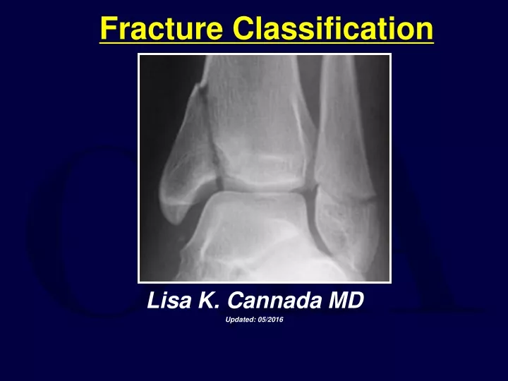
Fracture Classification Lisa K. Cannada MD Updated: 05/2016
History of Fracture Classification • 18th & 19th century • History based on clinical appearance of limb alone Colles Fracture Dinner Fork Deformity
20th Century • Classification based on radiographs of fractures • Many developed • Problems • Radiographic quality • Injury severity
What about CT scans? • CT scanning can assist with fracture classification • Example: Sanders classification of calcaneal fractures
The Soft Tissues Fracture appears non complex on radiographs The real injury
Patient Variables • Age • Gender • Diabetes • Infection • Smoking • Medications • Underlying physiology
Injury Variables • Severity • Energy of Injury • Morphology of the fracture • Bone loss • Blood supply • Location • Other injuries
Why Classify? • As a treatment guide • To assist with prognosis • To speak a common language with other surgeons
As a Treatment Guide • If the same bone is broken, the surgeon can use a standard treatment • PROBLEM: fracture personality and variation with equipment and experience
To Assist with Prognosis • You can tell the patient what to expect with the results • PROBLEM: Does not consider the soft tissues or other compounding factors
To Speak A Common Language • This will allow results to be compared • PROBLEM: Poor interobserver reliability with existing fracture classifications
Interobserver Reliability Different physicians agree on the classification of a fracture for a particular patient
Intraobserver Reliability For a given fracture, each physician should produce the same classification
Descriptive Classification Systems • Examples • Garden: femoral neck • Schatzker: Tibial plateau • Neer: Proximal Humerus • Lauge-Hansen: Ankle
Literature • 94 patients with ankle fractures • 4 observers • Classify according to Lauge Hansen and Weber • Evaluated the precision (observer’s agreement with each other) Thomsen et al, JBJS-Br, 1991
Literature • Acceptable reliabilty with both systems • Poor precision of staging, especialy PA injuries • Recommend: classification systems should have reliability analysis before used Thomsen et al, JBJS-Br, 1991
Literature • Classified identical 22/100 • Disagreement b/t displaced and non-displaced in 45 • Conclude poor ability to stage with this system • 100 femoral neck fractures • 8 observers • Garden’s classification Frandsen, JBJS-B, 1988
OTA Classification • There has been a need for an organized, systematic fracture classification • Goal: A comprehensive classification adaptable to the entire skeletal system! • Answer: OTA Comprehensive Classification of Long Bone Fractures
With a Universal Classification… To… Treatment Implant options Results You go from x-ray….
To Classify a Fracture • Which bone? • Where in the bone is the fracture? • Which type? • Which group? • Which subgroup?
Using the OTA Classification • Where in the bone? • Which bone?
Proximal & Distal Segment Fractures • Type A • Extra-articular • Type B • Partial articular • Type C • Complete disruption of the articular surface from the diaphysis
Diaphyseal Fractures • Type A • Simple fractures with two fragments • Type B • Wedge fractures • After reduced, length and alignment restored • Type C • Complex fractures with no contact between main fragments
Grouping-Type A • Spiral • Oblique • Transverse
Grouping-Type B • Spiral wedge • Bending wedge • Fragmented wedge
Grouping-Type C • Spiral multifragmentary wedge • Segmental • Irregular
Subgrouping • Differs from bone to bone • Depends on key features for any given bone and its classification • The purpose is to increase the precision of the classification
OTA Classification • It is an evolving system • Open for change when appropriate • Allows consistency in research • Builds a description of the fracture in an organized, easy to use manner
Classification of Soft Tissue Injury Associated with Fractures
Closed Fractures • Fracture is not exposed to the environment • All fractures have some degree of soft tissue injury • Commonly classified according to the Tscherne classification • Don’t underestimate the soft tissue injury as this affects treatment and outcome!
Closed Fracture Considerations • The energy of the injury • Degree of contamination • Patient factors • Additional injuries
Tscherne Classification • Grade 0 • Minimal soft tissue injury • Indirect injury • Grade 1 • Injury from within • Superficial contusions or abrasions
Tscherne Classification • Grade 2 • Direct injury • More extensive soft tissue injury with muscle contusion, skin abrasions • More severe bone injury (usually)
Tscherne Classification • Grade 3 • Severe injury to soft tisues • -degloving with destruction of subcutaneous tissue and muscle • Can include a compartment syndrome, vascular injury Closed tibia fracture Note periosteal stripping Compartment syndrome
Literature • Prospective study • Tibial shaft fractures treated by intramedullary nail • Open and closed • 100 patients Gaston, JBJS-B, 1999
Literature What predicts outcome? Classifications used: • AO • Gustilo • Tscherne • Winquist-Hansen (comminution) All x-rays reviewed by single physician Evaluated outcomes Union Additional surgery Infection Tscherne classification more predictive of outcome than others Gaston, JBJS-B, 1999
Open Fractures • A break in the skin and underlying soft tissue leading into or communicating with the fracture and its hematoma
Open Fractures • Gustilo-Anderson • OTA-Open Fracture Classification (OFC)
Open Fractures • Commonly described by the Gustilo system • Model is tibia fractures • Routinely applied to all types of open fractures • Gustilo emphasis on size of skin injury
Open Fractures • Gustilo classification used for prognosis • Fracture healing, infection and amputation rate correlate with the degree of soft tissue injury by Gustilo • Fractures should be classified in the operating room at the time of initial debridement • Evaluate periosteal stripping • Consider soft tissue injury
Type I Open Fractures • Inside-out injury • Clean wound • Minimal soft tissue damage • No significant periosteal stripping
Type II Open Fractures • Moderate soft tissue damage • Outside-in mechanism • Higher energy injury • Some necrotic muscle, some periosteal stripping
Type IIIA Open Fractures • High energy • Outside-in injury • Extensive muscle devitalization • Bone coverage with existing soft tissue not problematic Note Zone of Injury
Type IIIB Open Fractures • High energy • Outside in injury • Extensive muscle devitalization • Requires a local flap or free flap for bone coverage and soft tissue closure • Periosteal stripping
Type IIIC Open Fractures • High energy • Increased risk of amputation and infection • Major vascular injury requiring repair
Literature on Open Fracture Classification • 245 surgeons • 12 cases of open tibia fractures • Videos used • Various levels of training (residents to trauma attendings) Brumback et al, JBJS-A, 1994
Literature on Open Fracture Classification • Interobserver agreement poor • Range 42-94% for each fracture • Least experienced-59% agreement • Orthopaedic Trauma Fellowship trained-66% agreement Brumback et al, JBJS-A, 1994