Download
1 / 54
570 likes | 981 Views
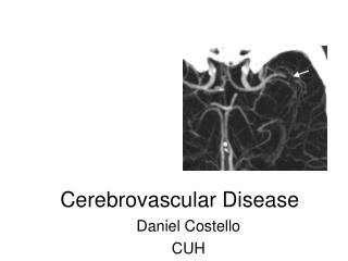
Cerebrovascular Disease. FM Brett , MD, FRCPath. ~ In USA 0.5 million new strokes diagnosed annually and 3 million survivors of a previous stroke. ~ Stroke 3 rd most frequent cause of death ~ 2 nd most frequent cause of dementia ~ Major reason for severe disability and long term

E N D
Cerebrovascular Disease FM Brett , MD, FRCPath
~ In USA 0.5 million new strokes diagnosed annually and 3 million survivors of a previous stroke ~ Stroke 3rd most frequent cause of death ~ 2nd most frequent cause of dementia ~ Major reason for severe disability and long term dependency
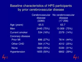
Epidemiological aspects of stroke ~ In the USA stroke is the third commonest cause of death ~ Incidence increases with age ~ Major risk factors for stroke are hypertension, cardiac disease, smoking, hyperlipidemia, and diabetes ~ Other causes OCP, sickle cell, coagulation disorders ~ In USA - brain infarction 10 times commoner than haemorrhage
Blood supply to the brain ~ Human brain approx 2% of body weight ~ Receives 15% of total cardiac output O2 consumption approximately 20% of whole body (i.e high metabolic rate) ~ How long would the brain survive if blood flow interrupted
Terminology ~ Ischaemia - arterial stenosis or occlusion Infarction - perfusion territory of the affected vessel ~ Global brain ischaemia - < CPP below the threshold for autoregulation i.e when systemic blood pressure falls very low e.g cardiac tamponade, heroin overdose, or ICP rises to a level that compromises cerebral perfusion Resultant brain damage or infarction is accentuated in the WATERSHED REGIONS
CPP= SAP - ICP CPP > 40 mmHg - necessary for autoregulation If CPP < 40 mmHg CBF falls dramatically
Principal causes of hypoxia Hypoxemic hypoxia - low O2 in blood ~ Carbon monoxide poisoning ~ Near drowning ~ Respiratory arrest ~ Prolonged status epilepticus Stagnant hypoxia - (inadequate supply of oxygenated O2 ~ Cardiac arrest ~ Rise ICP ~ Respirator brain Histotoxic hypoxia (inability of tissues to use O2) ~ Cyanide and sulphide exposure - inhibition of mitochondrial enzymes involved in oxidative respiration
Selectively vulnerable zones ~ Hippocampus - CA1 ~ Laminae 3 and 5 of cortex ~ Purkinje cells cerebellum
HYPOXIA- blood flow to the CNS may be normal or increased Damage occurs in selectively vulnerable neurones
Hypoxic ischaemic encephalopathy ~ Variable clinical presentation ~ Clinical recovery generally better after hypoxemic hypoxia than after global brain ischaemia ~ Severity and duration of HIE after transient cerebral hypoxia depends on I) duration of insult 2) completeness of insult 3) blood glucose level (high level poor outcome) 4) CNS temperature ~ Long term sequelae - difficult to predict
Infarct - region of cell death Infarct may become secondarily haemorrhagic and may mimic a primary haemorrhage STROKE - rapid onset of focal disturbance of cerebral function lasting > 24 hours TIA - less than 24 hours Infarcts may be caused by: ~ Large vessel or macrovasculature disease ~ Small vessel or microvasculature disease ~ Emboli ~ Venous thrombosis
Types of vascular disease Anterior circulation A. Small vessel disease – e.g microangiopathy B. Occlusive large vessel disease of pial arteries C. Occlusive large- vessel disease of brain supplying arteries in the neck D. Embolising heart disease including aortic plaques and R-L shunts
1 a, b – small vessel disease 2 a,b. Atherosclerotic or embolic occlusion of cerebellar artery 3 – intracranial thrombosis 4 – atheromatosis of vertebro-basilar artery 5 – embolus sticking in mid-basilar artery • In situ thrombosis of • basilar artery
Thalamic infarction Sensory or sensiomotor hemi-deficits with disassociated sensory loss, hemispasticity or even severe impairment of position sense; Segmental and focal dystonia without jerks and abnormal dystonic posture of affected hand
~ Large vessel disease includes atherosclerosis, fibromuscular dysplasia, arterial dissection, giant cell arteritis ATHEROSCLEROSIS IS THE COMMONEST OF THESE
ATHEROSCLEROSIS - leading vasculopathy producing brain infarcts. Affects intracranial and extracranial large vessels RISK FACTORS ~ Hyperlipidemia ~ Hypertension ~ Cigarette smoking ~ Obesity ~ Age ~ Sex
Small vessel disease includes cerebral vasulitides PACNS ~ Isolated granulomatous or primary angiitis of the CNS (PACNS)
PACNS ~ Recognised in the mid 1950’s ~ Diagnosis: ~ Clinical ~ Imaging ~ Biopsy
EMBOLIC DISEASE • Embolic stroke results when any solid material: • forms within the aterial circulation • is introduced into the arterial circulation • forms within the venous and has a conduit to the • arterial circulation ì.e right to left shunt • Resultant infarct is : • clinically abrupt • haemorrhagic
Autopsy of a stroke patient If infarction 1. Examine major cranial arteries i.e carotid and vertebral arteries in the neck 2. Carefully examine heart for: infective endocarditis valvular abnormalities septal defects
IF haemorrhage Look for evidence of: 1. Hypertension i.e cardiomegaly, LVH, nephrosclerosis 2. Neoplasia 3. Drug abuse 4. If dementia CAA
Blood in the cranial cavity • Source? • Spread • Occur? • Cause? • Sufficient to cause death
Intracranial haemorrhage • Extradural • Subdural • Subarachnoid • Intracerebral
SAH • Berry aneurysm • Infectious • Fusiform aneurysm • AVM • CAA
Berry aneurysms • Congenital • Risk of bleeding inc; • Hypertension • AVM • systemic vascular • disease • defects collagen • polcystic renal disease
ICH causes • Hypertension • Trauma • CAA • Berry aneurysm • AVM • Bleeding diathesis • Vasculitides • Drugs • Neoplasm • Infective
Hypertension - major risk factor for brain haemorrhage • Occurs due to rupture of arterioles that have become • weakened • DUE TO • Replacement of smooth muscle by fibrocartilagenous material • Fragmentation of elastic tissue • Charcot-Bouchard aneurysms
Haemorrhages involving the basal ganglia- putamen in particular tend to be non-traumatic and caused by hypertension
Chronic hypertension leads to arteriolar sclerosis resulting in small lacunar infarcts OCCUR~ BASAL GANGLIA~ PONS~ DEEP WHITE MATTER
AVM • Commonest 3-4th decade • Rarely familial • Rarely multiple • have a nidus • commonest vascular malformation • identified in surgical specimens
End result of herniation is compression and Duret haemorrhages as seen in the pons
Venous Thrombosis ~ Often secondary to infectious causes ~ Pregnancy ~ Puerperium ~ OCP ~ Haematological abnormalities