Download

1 / 13
130 likes | 282 Views
Circulating Osteogenic Precursor cells in End-stage aortic valvular disease. Robert Pignolo Kevin Egan. Aortic valve stenosis is a pathogenic process of inflammation, calcification, and ectopic bone formation.

E N D
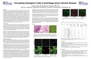
Circulating Osteogenic Precursor cells in End-stage aortic valvular disease Robert Pignolo Kevin Egan
Aortic valve stenosis is a pathogenic process of inflammation, calcification, and ectopic bone formation. Recently described COP (Circulating Osteogenic Precursor) cells are found in pre-osseous lesions of ectopic bone
Hypothesis: COP cells are responsible for the ectopic bone formation in end-stage valvular disease Are capable of inducing bone formation in muring inplanatation assays Can seed into inflammatory sites of tissue injury
Archived tissue samples of excised heart valves were evaluated for pathological bone formation
Heart Valve samples containing bone were stained for the presence of COP cells by looking for coexpression of type 1 collagen and CD45 (A) A stenotic heart valve with mature bone elements, including osteocytes and bone lining cells (arrow heads). In regions of fibroproliferation and neovascularity (B), COP cells were identified by CD45 (red) and Type 1 Collagen (green) staining (C). Original magnification 200X. A and B, Hemotoxylin & Eosin staining; C, Immunofluorescence of specific markers as described in Materials & Methods.
Each leaflet was arbitrarily divided into 4 regions and was histologically evaluated for the presence of bone. Regions without bone could serve as an internal negative control in addition to using pre-immune serum
Infiltration Fibroproliferation Neovascularity Cartilage Bone Bone Masson Trichrome of heart valve
Infiltration Fibroproliferation Neovascularity Bone Bone Cartilage Alizarin Red staining of heart valve
Picosirius Red staining of heart valve Fibroproliferation Neovascularity Cartilage Bone
In Vitro BMP staining of cultured COP cells BMP 4 BMP 2
IHC staining of heart valve CD45 Osteocalcin