Download
1 / 7
0 likes | 7 Views
This paper aims to examine the isolation of Lactobacillus from dairy products (milk, curd, and yogurt), extraction of Bacteriocin from it as well as to determine their inhibitory effect against few fungal skin pathogens and bacterial skin pathogens such as: Candida albicans, Aspergillus sp., Malassezia sp., Fusarium sp., and Penicillium sp., Staphylococcus aureus, Streptococcus pyogens, Klebsiella sp. The antagonistic activity of Lactobacillus sp. is mainly due to the bacteriocin present in it, therefore in this study,

E N D
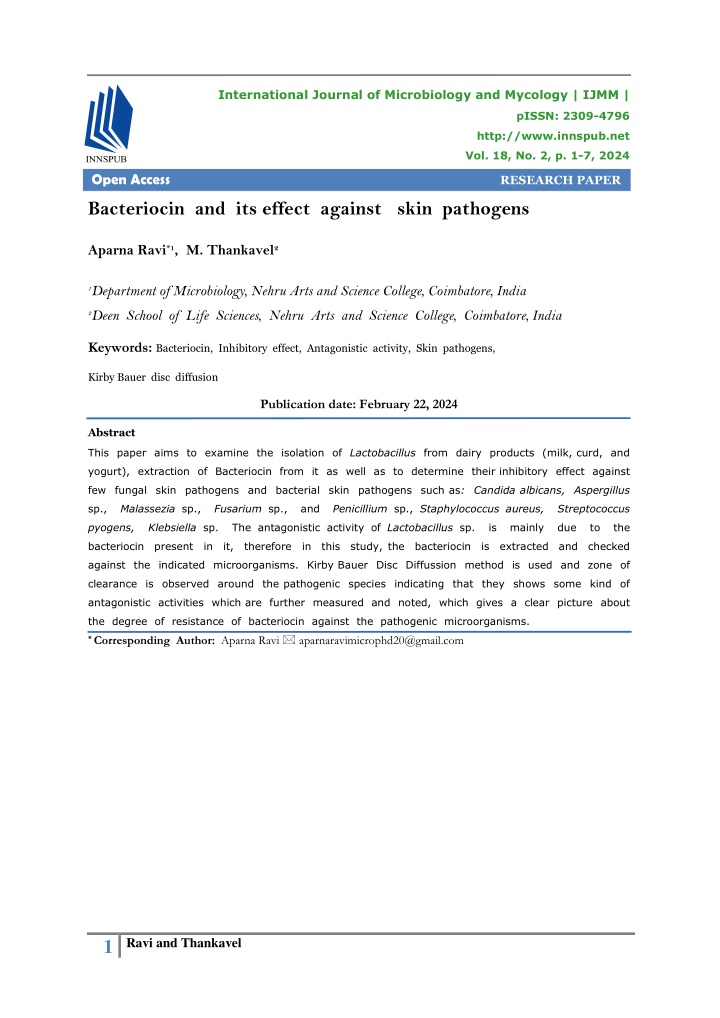
International Journal of Microbiology and Mycology | IJMM | pISSN: 2309-4796 http://www.innspub.net Vol. 18, No. 2, p. 1-7, 2024 Open Access RESEARCH PAPER Bacteriocin and its effect against skin pathogens Aparna Ravi*1, M. Thankavel2 1Department of Microbiology, Nehru Arts and Science College, Coimbatore, India 2Deen School of Life Sciences, Nehru Arts and Science College, Coimbatore, India Keywords: Bacteriocin, Inhibitory effect, Antagonistic activity, Skin pathogens, Kirby Bauer disc diffusion Publication date: February 22, 2024 Abstract This paper aims to examine the isolation of Lactobacillus from dairy products (milk, curd, and yogurt), extraction of Bacteriocin from it as well as to determine their inhibitory effect against few fungal skin pathogens and bacterial skin pathogens such as: Candida albicans, Aspergillus sp., Malassezia sp., Fusarium sp., and Penicillium sp., Staphylococcus aureus, Streptococcus pyogens, Klebsiella sp. The antagonistic activity of Lactobacillus sp. is mainly due to the bacteriocin present in it, therefore in this study, the bacteriocin is extracted and checked against the indicated microorganisms. Kirby Bauer Disc Diffussion method is used and zone of clearance is observed around the pathogenic species indicating that they shows some kind of antagonistic activities which are further measured and noted, which gives a clear picture about the degree of resistance of bacteriocin against the pathogenic microorganisms. * Corresponding Author: Aparna Ravi aparnaravimicrophd20@gmail.com 1 Ravi and Thankavel
Balanced skin is crucial for maintaining healthy Introduction skin functioning; but changes in the skin Skin is an important organ that represents the microbes are associated with skin diseases such first line of defense against the external as those caused by Candida albicans, Aspergillus, environment. Some microorganisms are Malassezia furfur, Fusarium, Penicillium, pathogenic but some are usually present on the Staphylococcus aureus, Streptococcus pyogens, skin does not causing damages but during and Klebsiella sp. Lactobacilli are probiotic adverse condition like immunosuppressant belonging to the group of lactic acid bacteria; phases the organisms develop infections in the they are Gram-positive, non-sporulating, host (Hall and Dorsch, 2002). Some of such cases anaerobic or facultative anaerobic rods. They are like; Primary cutaneous aspergillosis usually commonly present in dairy products, soil, lakes, involves site of injury, at or near intravenous and the intestinal tract of humans and animals. catheter sites, occlusive dressings, burns, or They possess antagonistic activity against various surgery (Walsh and Groll, 1999). Secondary pathogenic microorganisms (Salminen et al., infections infect the underlying structures or from 2004). In the present study, the Lactobacillus sp. wide spread blood borne seeding of the skin. from dairy products like milk, curd, and yogurt Superficial infections including keratitis, are isolated; then bacteriocin is extracted from it otomycosis are commonly caused by Penicillium and checked their efficacy against fungal and sp., Malassezia sp. are another example of bacterial pathogens using the technique of Kirby normal skin flora colonizes as commensals and Bauer disc diffusion method (Aasen and Moretro, during adverse conditions develops diseases at 2018). Bacteriocins are low molecular weight the head, neck dermatitis, and malassezia peptides secreted by the bacterial cells to kill folliculitis (Gupta and Kohli, 2004). sensitive cells present in the same ecosystem competing for food and other nutrients. Fusarium sp. Causes infections in patients in Bacteriocins, along with their native antibacterial conditions like hematologic malignant or bone property, also exhibit additional antiviral and marrow transplant (Nucci and Anaissie, 2007). antifungal properties nowadays (Riley and Wertz, Candida nail infections occur in patients with 2002). The dairy samples (milk, curd, yogurt) chronic mucocutaneous candidiasis caused by were collected in sterile containers from different Candida albicans, they invade the entire nail places of Vandithavalam, Palakkad, Kerala. The plates (Kirkpatrick, 2001). Staphylococcus aureus samples were analyzed microbiologically, is a major cause of bacterial skin infections identified, and confirmed by biochemical tests. namely, abscesses in boils, furuncles, Cellulitis The study aimed to extract bacteriocin from (Prendiville, 1989). Streptococcus pyogens are Lactobacillus sp. and to see whether its effect also bacterial species causing infections in the against the mentioned microorganism. superficial keratin layer called impetigo, the superficial epidermis layer-erysipelas, the Materials and methods subcutaneous tissue layer-cellulitis, fascia called Collection of milk samples necrotizing fasciitis or in the muscle-myositis, Raw milk samples of cow and goat were myonecrosis (Stevens and Bryant, 2017). collected in a sterile container in the month of February 2022 at various places of Klebsiella causes surgical wound infections, they Vandithavalam, Palakkad, Kerala for the usually enters through break in the skin and isolation of Lactobacillus species and gradually leads to soft tissue infections examined under aseptic conditions. (Paterson and Bonomo, 2005). 2 Ravi and Thankavel
Collection of curd samples Klebsiella Homemade curd samples and commercially Swabs were taken from patients with boils available curd samples of two different brands and wound with discoloration. were collected for the isolation of Lactobacillus sp. Isolation and characterization of Lactobacillus from dairy samples Collection of Yogurt samples 1.The milk, curd and yogurt samples were Yogurt samples of two different commercial serially diluted in various test tubes. brands were collected for the isolation of 2.MRS (de Man, Rogosa, and Sharpe agar- Lactobacillus sp. selective media for LB) agar plates were prepared. Collection of microorganisms 3.Swabs from various dilutions from each of Candida albicans the samples of milk, curd and yogurt were Swabs were taken from patients with toe swabbed onto MRS agar plates using sterile nail and skin infections characterized by fluid techniques and control plates were discharge , Colour changes of the nail, Pain in maintained. the nail and toe. 4.All the inoculated plates of three different samples of different dilutions were incubated Malassezia at 37 degree Celsius for exactly 24 hours Malassezia samples were collected by taking and then plates were observed for growth. the Scrappings from various patients of head 5.Colonies formed in the plates were and neck dermatitis characterized by white patchy scales present in the affected areas subjected for Staining and biochemical tests with itching and dryness. for the confirmation of the species of microorganisms present. Aspergillus Swabs were collected from patients with skin Grams staining injury and burns. A clean grease free glass slide was taken, washed thoroughly, surface sterilised using Fusarium ethanol and smear of single colony has been Swabs were collected from skin lesions through made on the slides and allowed to dry, the aseptic techniques. smear is heat fixed by passing the slide above flame of Bunsen burner for 2-3 times. Penicillium The smear was cooled, flooded with the Swabs were taken from patients of skin primary stain -crystal violet, then waited for lesions with swelling and redness in the skin one minute was washed it under running with discolouration. tap water. Then added the Mordant- Gram’s iodine, waited for one minute, decolorized it with Staphylococcus aureus 95% ethyl alcohol, washed under running tap Swabs were taken from the pimples of water. Finally, added few drops of the patients with granules and pus. counter stain-Safranin, waited for one minute and washed under running tap water, air Streptococcus pyogens dried and observed under oil immersion of Discharges from the wound is collected 100X (Buchanan, 1982). through sterile cotton swabs. 3 Ravi and Thankavel
Catalase test sliding above a glass rod. Then streaked with A clean glass slide was taken, washed and the test organism and incubated at 37°C for 24 hours(Schlegel, 1976). added a drop of hydrogen peroxide to the center of the slide, then fresh culture of test organism is mixed properly to it using a tooth Carbohydrate fermentation test pick or inoculation loop and observed for the presence of bubble formation(Schlegel, 1976). Glucose, Fructose, Sucrose, Xylose, Lactose fermentations were checked. The test was performed by using 1% sugar in MRS broth. Oxidase test Media was prepared and poured into test tubes Oxidase disc was placed in a moisture free and Durham’s tube was inserted invertably area and a small portion of test culture is into it. Test culture is inoculated and the spreaded on to it using a toothpick or tubes were incubated at 37°C for 24 hours. inoculation loop, observed for the presence of colour change(Beveridge, 2001). Phenol red was used as an indicator and observed the colour changes and formation of gas bubbles(Buchanan, 1982). Indole test Tryptophan broth was prepared, poured into Extraction of Bacteriocin test tubes, sterilized, cooled and inoculated Extraction of bacteriocins from Lactobacillus was method with the test organisms, incubated at 37°C done using chloroform extraction for 24 hours. After incubation few drops of (Beveridge, 2001). Kovac’s reagent as an indicator was added to the test tubes and observed the colour change(Buchanan, 1982). Isolation of microorganism Candida albicans Sabouraud Dextrose Agar (SDA) media was Methyl red test prepared and the Samples collected from patients MR-VP broth was prepared, poured into sterile were swabbed onto it. Plates were incubated at 37°C for 2-5 days and growth was observed test tubes and sterilized. Broth was inoculated with the test organisms and incubated at 37°C (Buchanan, 1982). for 24 hours. After incubation Methyl red reagent was added and observed the colour change(Beveridge, 2001). Malassezia Dandruff like flaky scrapings from patients were introduced to Dixon agar and SDA agar Voges-Proskauer test plates and incubated at 37°C for 3-5 days and observed growth(Buchanan, 1982).. MR-VP broth was prepared and poured into sterile test tubes and sterilized, inoculated with the test organism and incubated at 37°C Aspergillus, Penicillium and Fusarium for 24 hours. After incubation few drops of Swabs taken from patients were introduced Barrit’s reagent A and Barrit’s reagent B, is to SDA plates and incubated at 35°C for 2-5 days(Buchanan, 1982). added, Shaked well and observed the colour change(Buchanan, 1982). Staphylococcus aureus Citrate utilization test Swabs from patients were inoculated into Simmon citrate agar medium was prepared and HiCrome Staph Selective Agar plates and sterilised, poured into test tubes and slants incubated at 37°C for 24 hours (Buchanan, were prepared by keeping the test tubes by 1982). 4 Ravi and Thankavel
Streptococcus pyogens Table 1. Morphological characteristics of Swabs from patients were inoculated into Lactobacillus Nutrient agar plates, incubated at 37°C for 24 hours(Buchanan, 1982). Characteristics LB -1 LB -2 observations Creamy pale white colonies were seen Pale white coloured colonies were seen Purple coloured rods in chains were observed Observations Small white raised colonies were seen Pale white coloured round colonies were seen Purple coloured rods in chains were observed Colony morphology on Nutrient agar Klebsiella Colony morphology on MRS agar Swabs from patients were inoculated into MacConkey agar plates, incubated at 37°C for 24 hours (Buchanan, 1982). Gram staining Checking the antagonistic activity Antimicrobial activity was tested by disc diffusion method(Beveridge, 2001). Candida albicans, Table 2. Biochemical characteristics of Lactobacillus Aspergillus, Malassezia, Fusarium and Penicillum, Name of tests Indole Methyl red Voges- proskauer Citrate utilization Oxidase Catalase Triple sugar iron agar Results (LB-1) Positive Positive negative Results (LB-2) Positive Positive negative Staphylococcus aureus, Streptococcus pyogens and Klebsiella samples were swabbed onto separate Muller Hinton Agar plates, sterile discs coated with bacteriocins were placed on it. Positive Positive The plates were incubated and observed the zone Negative Positive Negative Negative Positive Negative of inhibition around the discs. The diameter of zone was measured. Results Glucose positive Lactose positive Sucrose positive Glucose positive Lactose positive Sucrose positive Carbohydrate utilisation In the MRS agar plates, small to medium white colonies were observed. Gram staining Table 3. Measurement of zone of inhibition showed purple rods indicating Gram-positive rod against bacteriocin of Lactobacillus shaped bacteria (Fig. 1, Table 1). Organisms Candica albicans Aspergillus sp. Malassezia furfur Fusarium sp. Penicillium Staphylococcus aureus Streptococcus pyogens Klebsiella Diameter of zone (mm) 14mm 15mm 14.5mm 13mm 12mm 12.5mm 13mm 13mm Biochemical tests showing: oxidase- positive, catalase- positive, Indole -positive, MR -positive, Citrate- positive, lactose fermentation- positive indicated that the isolated microorganism is confirmed as the (LAB) Lactic acid bacteria- Lactobacillus sp. (Table 2). Bacitracin was extracted from the organism. Candida albicans, Aspergillus, Fusarium and Penicillum, Staphylococcus aureus, Streptococcus pyogens, Klebsiella swabbed plates were inoculated with the bacteriocin Colonies on MRS Agar Malassezia, Microscopic observation of LB Fig. 1. Colonies on MRS Agar and microscopic observation of LB 5 Ravi and Thankavel
confirmed that it has the ability to kill or suppress coated discs, zone of clearance was observed. The diameter of zone was measured and tabulated (Fig. 2, Table 3). the growth of various fungi and bacteria (Cotter et al., 2005). Antagonistic activity of Bacteriocin of Lactobacillus sp isolated from dairy samples was subjected to the study and observations against fungal and bacterial pathogens, showing good degrees of inhibition (Mokoena, 2017). Considering this as a reference, recent studies are ongoing, expecting the formulation of new Candida sp. Fusarium sp. therapeutics from bacteriocins, which could be a broad-spectrum drug and a solution for the challenge of multidrug resistance shown by various pathogens (Perez et al., 2014). Conclusion Aspergillus sp. Malasseziasp. A total of two raw milk samples from cow and two from goat, two curd samples (homemade, commercially available), two yogurt samples were collected, Morphological, physiological, microscopic observation, Penicillium sp. Streptococcus sp. biochemical characterisations were made and confirmed as Lactobacillus species. The antagonistic activity of bacteriocin is checked. They are found to be sensitive against human pathogen Candida albicans, Aspergillus sp., Malassezia sp., Fusarium sp., Penicillium S. aureus Fig. 2. Zone of inhibition against bacteriocin of Lactobacillus Klebsiella sp. sp., Staphylococcus aureus, Streptococcus pyogens, Klebsiella sp. causing various skin Discussion infections in human beings. This has an important role in medical microbiology and One of the major challenges that microbiologists human health. are facing nowadays is the multidrug resistance shown by various pathogenic species (Ventola, References 2015). If the compound from a microorganism Aasen IM, Moretro T. 2018. Extraction and itself is efficient to control the growth and Purification of Bacteriocins from Lactic Acid adverse effects of pathogens, it will be a Bacteria. In Bacteriocins: Production, Applications revolutionary moment (Fernández, 2019). The and Safety (pp. 73-91). Springer. use of probiotics is one example of such an application. Probiotics have been considered Beveridge TJ. 2001. Use of the Gram stain in effective in maintaining gut health for many microbiology. Biotechnic & Histochemistry 76(3), years, and now they are being explored for more 111-118. possible applications (Hill et al., 2014). The probiotic Lactic acid bacteria, Lactobacillus, is Buchanan RE. 1982. Microbial staining methods. isolated, and its virulent compound, Bacteriocin, ASM Press. is extracted, purified, and checked. It is 6 Ravi and Thankavel
Cotter PD, Hill C, Ross RP. 2005. Bacteriocins: Paterson DL, Bonomo RA. 2005.Extended- spectrum beta-lactamases: a clinical update. Clin Developing innate immunity for food. Nature Microbiol Rev. 18(4), 657-86. Reviews Microbiology 3(10), 777-88. DOI: 10.1128/CMR.18.4.657-686.2005. DOI: 10.1038/nrmicro1273. Perez RH, Zendo T, Sonomoto K. 2014. Novel Fernández L. 2019. Fighting Fire with Fire: bacteriocins from lactic acid bacteria (LAB): Exploiting Bacterial Antagonism to Combat Drug various structures and applications. Microbial Cell Factories 13(Suppl 1), S3. Resistance. Trends in Microbiology 27(4), 168- 174. DOI: 10.1186/1475-2859-13-S1-S3 Prendiville JS, Hebert AA, Greenwald MJ, Esterly NB. 1989. Management of Stevens- Johnson syndrome and toxic epidermal necrolysis in children. J. Pediatr. 115(6), 881-7. DOI: 10.1016/s0022-3476(89)80736-x. Riley MA, Wertz JE. 2002. Bacteriocins: Evolution, Ecology, and Application. Annual Review of Microbiology 56, 117-137. DOI: 10.1146/annurev.micro.56.012302.161024. Salminen S, von Wright A, Ouwehand A. 2004. Lactic Acid Bacteria: Microbiological and Functional Aspects (4th ed.). CRC Press. https://doi.org/10.1201/b11503 Gupta AK, Kohli Y. 2004.Prevalence of Malassezia species on various body sites in clinically healthy subjects representing different age groups. Med Mycol. 42(1), 35-42. DOI: 10.1080/13693780310001610056. Hall GS, Dorsch MM. 2002. Skin and soft tissue infections. In Principles and Practice of Infectious Diseases (6th ed., Vol. 1, pp. 309-331). Elsevier. Hill C, Guarner F, Reid G, Gibson GR, Merenstein DJ, Pot B, Sanders ME. 2014. Expert consensus document: The International Scientific Association for Probiotics and Prebiotics consensus statement on the scope and Schlegel Cambridge University Press. appropriate use of the term probiotic. Nature HG. 1976. General Microbiology. Reviews Gastroenterology & Hepatology 11(8), 506-514. DOI: 10.1038/nrgastro.2014.66. Stevens DL, Bryant AE. 2017.Necrotizing Soft- Tissue Infections. N. Engl. J. Med. 377(23), 2253-2265. DOI: 10.1056/NEJMra1600673. Kirkpatrick CH. 2001.Chronic mucocutaneous candidiasis. Pediatr Infect Dis J. 20(2), 197-206. Ventola CL. 2015. The antibiotic resistance crisis: part 1: causes and threats. P&T: A Peer- Reviewed Journal for Formulary Management 40(4), 277-283. DOI: 10.1097/00006454-200102000-00017. Mokoena MP. 2017. Lactic acid bacteria and their bacteriocins: classification, biosynthesis and applications against uropathogens: a mini-review. Walsh TJ, Groll AH. 1999.Emerging fungal Molecules 22(8), 1255. pathogens: evolving challenges to Nucci M, Anaissie E. 2007.Fusarium infections in immunocompromised patients for the twenty-first immunocompromised patients. Clin Microbiol Rev. century. Transpl Infect Dis. 1(4), 247-61. 20(4), 695-704. DOI: 10.1128/CMR.00014-07. DOI: 10.1034/j.1399-3062.1999.010404.x. 7 Ravi and Thankavel