Download
1 / 17
0 likes | 11 Views
Highly diverse biological system of solar salterns with different salinities, often provide high densities of mycofloral populations, makes the salterns excellent model systems for both its diverse and activity. In this study, diversity of halophilic fungi in six stations which includes reservoir, evaporator and crystallizer pond of both Marakkanam and Tuticorin saltpans in relation to environmental parameters were carried out for a period of two years. 95 species of halophilic fungi from water and sediment samples belongs to 41 genera were recorded in both saltpans.

E N D
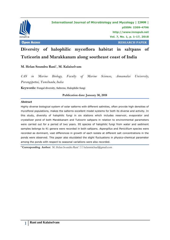
International Journal of Microbiology and Mycology | IJMM | pISSN: 2309-4796 http://www.innspub.net Vol. 7, No. 1, p. 1-17, 2018 Open Access RESEARCH PAPER Diversity of halophilic mycoflora habitat in saltpans of Tuticorin and Marakkanam along southeast coast of India M. Helan Soundra Rani*, M. Kalaiselvam CAS in Marine Biology, Faculty of Marine Sciences, Annamalai University, Parangipettai, Tamilnadu, India Keywords: Fungal diversity, Salterns, Halophilic fungi Publication date: January 30, 2018 Abstract Highly diverse biological system of solar salterns with different salinities, often provide high densities of mycofloral populations, makes the salterns excellent model systems for both its diverse and activity. In this study, diversity of halophilic fungi in six stations which includes reservoir, evaporator and crystallizer pond of both Marakkanam and Tuticorin saltpans in relation to environmental parameters were carried out for a period of two years. 95 species of halophilic fungi from water and sediment samples belongs to 41 genera were recorded in both saltpans. Aspergillus and Penicillium species were recorded as dominant, vast differences in growth of each isolate at different salt concentrations in the ponds were observed. This paper also elucidated the slight fluctuations in physico-chemical parameter among the ponds with respect to seasonal variations were also recorded. * Corresponding Author: M. Helan Soundra Rani* helanmichael@gmail.com 1 Rani and Kalaiselvam
Introduction Enumeration of fungi in these habitats revealed Saltpans are man-made seasonal ponds their presence in relatively large numbers (up to constructed mainly for the production of raw salt. 4×10⁴ ml–1), but the biodiversity appears to be These ponds offer an experimental system with limited to a small number of fungal genera. At an extreme environmental conditions include high present, 106 orders of fungi were known to and low temperature, pH, pressure, salt tolerate at low water activity(Kirk et al., 2001). concentration, low nutrient concentration, water Within Ascomycota, the main orders with halophilic availability and also conditions having high levels and halotolerant representatives are Capnodiales, of radiation, harmful heavy metals, toxic Sporidiales, Dothideales and Eurotiales. Both compounds (organic solvents) and strong orders Capnodiales and Dothideales have a gradient in biodiversity of primary and secondary xerotolerant tendency, as they contain a large producers. (Satyanarayana et al., 2005). It is one number of extremotolerant species that can grow such example for thalassohaline environment, it as epilithic or cryptoendolithic species at high or contains the salinity range of five to ten times low temperatures (Selbmann et al., 2005) and saltier than seawater (150-300 g/l salt hypersaline coastal areas worldwide. concentration). Life at high salt concentrations This new ecological findings are not only important requires special adaptations of the cell’s for our understanding of microbial processes in physiology. Microbes must sense environmental hypersaline environments worldwide, but also for stresses, transduce these signals and mount not yet fully acknowledged. Though, the sequence protective responses to survive in hostile of works regarding halophilic fungi from solar environments (Nikolaou et al., 2009). saltern environments has been carried out for the past two decades in many continents but the Most microbial diversity studies in salterns have meager works were contributed by Indian focused on halophilic Archaea bacteria of the order Halobacteriales, which comprise the main subcontinent. Owing to the lack of studies on microbial component in these environments mycofloral in salterns along the Indian coast, the (Oren, 2002). Other organisms such as algae, present study was carried out to understand the protozoa, eubacteria and even fungi are also ecology and diversity, seasonal variations, found in the salterns, even though it was thought frequency of occurrence and distribution of fungi in that they could not survive under extreme salt relation to physico-chemical parameters in conditions (Gunde-Cimerman et al., 2004). Fungi Tuticorin and Marakkanam saltpans along are ubiquitous in most ecosystems where they southeast coast of India. usually colonize a diverse range of substrates. Materials and methods Fungal cell adaptations to high saline Monthly samples were made to record the environment are the promising biological process physico-chemical parameters and to check the and the level of plasma-membrane fluid fungal diversity. Rainfall data was obtained from fluctuation are indicators of fitness for survival the local meteorological unit under government of and adaptability in fungi obtained from extreme India located at Villupuram and Tuticorin. Field environments (Turk et al., 2007). Unique in-situ data like temperature, salinity, dissolved oxygen morphology was interpreted as a response to and pH were measured. Atmospheric and surface multiple stress factors which can adapt to water temperatures were measured using extreme conditions. The accumulation of standard mercury filled centigrade thermometer. osmoprotective compounds such as polyols Salinity was estimated by hand refractometer (glycerol) sugars (trehalose and manitol) and (Atago, Japan) and pH was measured using Elico some unusual amino acids may also play an pH meter (Model LC- 120). important role under salt stress (Griffith, 1994). 2 Rani and Kalaiselvam
Dissolved oxygen was estimated by the modified programme. Origin 8 and PRIMER (Ver-6) were sed Winkler’s method (Strickland and Parsons, 1972). for univariate and multivariate analyses. For the analysis of nutrients, surface water Periodicity of occurrence samples were collected in clean polyethylene It denotes the number of samplings in which a bottles and kept in an ice box and transported to particular fungus was recorded against the total the laboratory. The water samples were filtered number of samplings. Based on the percentage using a Millipore filtering system and analyzed for periodicity of occurrence, fungi were classified dissolved inorganic phosphate, nitrate and into four groups as Most common (76-100%), reactive silicate. Sediment samples were dried in Common (51-75%), Occasional (26-50%) and an oven at 105°C for 6 hours and ground using Sporadic (1-25%). mortar and pestle for breaking and were sieved before analysis. Sediment nutrients like Phosphate (TP), Nitrogen (TN) and Total Organic Carbon (TOC) In the present study, the data were analyzed for by adopting the standard methods described by diversity index (H’) using the following Shannon- Strickland and Parsons (1972). Wiener’s formula (1949) A calendar year was divided into four seasons Species richness viz., monsoon (October to December), post Species richness (d) was calculated using formula monsoon (January to March), summer (April to given by Margalef June) and pre-monsoon (July to September) based on the north east monsoon which is prevalent in the study area. Pielou’s evenness index The fungal strains from water and sediment The equitability (J’) was computed using the following formula of Pielou (1996): samples of all the six stations were isolated using standard pour plate technique. In mycological analysis, water and sediment samples were Graphical/ distributional methods plated on to Potato Dextrose Agar + 20% solar K-dominace plot salt for isolation of halophilic fungi, which were Dominance plot is also called as the ranked then incubated at room temperature of about species abundance plot developed by Lambshead 30°C, up to 15 days. et al. (1983). The isolates were picked based on dissimilarity in the colony characteristics, purified and numbered Ellipse plot according to the stations and the samples. Fungal Average taxonomic distinctness index (delta+) identification was done on the basis of colony and and variation in taxonomic distinctness microscopic morphology characteristics with (lambda+) were studied graphically. Being based reference to identification keys (Ellis, 1971). on the presence or absence of species according Based on the results, statistical analyses were to seasonal variations, they can be calculated assist to make the cleared data output. Statistical Analysis using simple species list. Average taxonomic distinctness (∆+) and Further, various statistical methods such as variation in taxonomic distinctness (λ+) were Univariate, graphical/distributional and multivariate calculated for all staions during all the seasons were applied for the data analysis. The computer from the study area. 3 Rani and Kalaiselvam
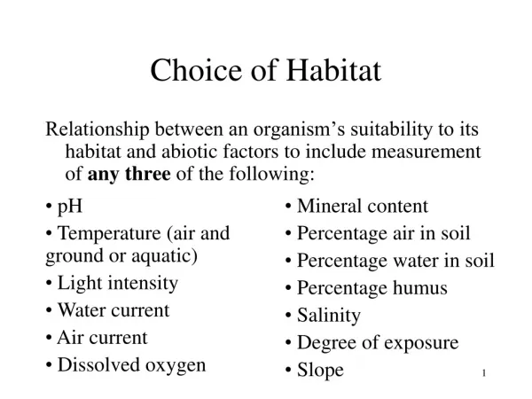
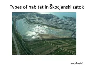
Study area Fig. 1. Map showing the sampling areas in Marakkanam and Tuticorin, India. Results and discussion The physico-chemical parameters and nutrients The pH of brine at different stages of the salt pan of both water and sediment samples in Tuticorin was alkaline in nature (Baati et al., 2008). In the and Marakkanam saltpans showed the variations present study, the pH encountered high during in all the six stations. In the present study, brine summer and minimum in monsoon season temperatures at different stations vary, showed the moderate ranges between 7-7.8 maximum temperature was observed during (alkaline) in both Tuticorin and Marakkanam summer due to the intensity of solar radiation salterns. This study was highly supported by heat the brine water of shallow ponds of salterns Manikandan et al. (2009) and Radhika et al. resulting in rapid evaporation of water causing (2011), who observed the range from 7.35- 7.85 speedy crystallization process. In case of in Marakkanam salterns and 7.1-8.3 in Tuticorin sediment, minimum temperature was recorded salterns respectively. Butinar et al. (2005a) compared with water. Slight increase in brine highlighted that pH in all the ponds were temperature was observed from reservoir to approximately low (7.2) from the beginning of crystallizer ponds, which was highly collaborated December to the mid of February. Due to the with the study of Petrovic (1998). This might be uptake of CO2 by the photosynthesizing due to the red pigmentation produced by organisms, especially phytoplankton and phototrophic microorganisms and the planktonic cyanobacteria from the seawater could photosynthetic primary producer in crystallizer have increased the pH level during the summer ponds that cause β-carotene accumulation by the season (Subramanian and Mahadevan, 1999). green alga Dunaliella salina which was the main The low pH observed during the monsoon due to or sole primary producer in these ponds the influence of freshwater influx and dilution of (Litchfield, 1991). This red pigmentation brines, reduction of salinity and temperature and increases light absorption by the brine and decomposition of organic matter (Subramanian increases its temperature, thus enhancing the and Kannan, 1998). Gradual decrease of pH (7.1- salt production process (Javor, 2002). 7.65) from station-1 to station-3 was noted. 4 Rani and Kalaiselvam
Femitha and Vaithyanathan (2012) observed the Finally, to the crystallizer pond where the salinity pH value increases from source (seawater) to maintained up to 300-600‰, in this water masses reservoir because of the increasing concentration the salinity is considerably high and never gone of iron oxide and calcium carbonate but there below 250‰. This might be due to the water was a marginal decrease in the values at evaporates, gypsum and other minerals precipitate, evaporator and crystallizer stages because of the eventually, sodium chloride (NaCl) precipitates and removal of above mentioned salts before the salinity increases above 300 psu (Oren, 2002). brine pass on to the evaporator ponds. The soil samples were slightly acidic to neutral and the pH Lower silicate was recorded during summer season, ranged from 6.9-8.3 reported by Biswas and Paul while it was higher in monsoon period. The study (2012). Nazareth et al. (2012) stated that the pH was supported by Kovac (2009) reported that lower of sediments in the range of 5.3. concentration of silicate during summer, due to the considerable reduction in the freshwater input and In this study, salinity fluctuated widely between greater utilization of silicate by the abundantly stations. It went up high due to excessive occurring phytoplankton and cyanobacteria for their evaporation in the summer period and dropped biological activity (Nedumaran and Perumal, 2012). sharply during the monsoon season, the brine Nitrate concentrations were high (5.51μmol l−1) in salinity ranged between 395-400‰ and 400- crystallizer ponds and evaporator ponds ranked 410‰ in case of saline soils during summer second during monsoon season. Madkour and seasons of both salterns of Tuticorin and Gaballah (2012) recorded the data which supported Marakkanam. Salinity in these ponds reaches as the study and reasoned, organic and inorganic high as 400‰ during the peak salt producing nutrients caused hyper-nitrification, possibly season (summer) and as low as 5‰ during the phytoplankton blooms, oxygen depletion and monsoons (Kamat and Kerkar, 2011). formation of anoxie situation which was observed in Comparatively, the salinity showed vast variation pond (Ehrlich, 1987). Anbazhagan (1988) have among brine and saline soils this could be due to suggested that the addition of nitrogenous nutrients precipitation of halites from the water column due mainly through freshwaters and terrestrial runoff in to evaporation could be responsible for the higher the lagoon definitely increase the levels of nitrate. salinity of the sediment as compared to brine Lower concentration of nitrate was recorded which (Oren, 2003). Pedros-Alio et al. (2000) observed may be due to the utilization of benthic algae and that as temperature increases, salinity also phytoplankton. Inorganic phosphate concentration increase due to the higher calorific capacity of the showed slight variation from reservoir to crystallizer brines. Gradual increase of salinity was noted ponds which may be due to decomposition of from reservoir to crystallizer ponds, which might bacteria and further brine concentration (Dundas be due to the initial sea water pumped from the and Halvorson, 1966). ocean directly, from there the water almost continuously flows into other ponds and so the O2 are often short supply in hypersaline salinity ranged not much difference from the environments, because of its limited solubility at adjacent inshore waters of the sea and the initial high salt concentration (Nealson and Stahl, 1997) reservoir pond, the salinity ranged 35- 37‰. In and other point of interest were under extreme second category, the water was allowed to pass conditions O2 or organic nutrients become on to the evaporator pond where the water was limiting, thus making this nutritional extremophily stored for some days for evaporation enhanced as a commonly encountered adaptation in harsh by strong local winds and temperatures and the environments. In this present survey, oxygen salinity increases reached up to 100-200‰. values in the first pond reached as high and 5 Rani and Kalaiselvam
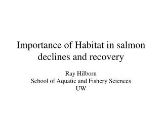
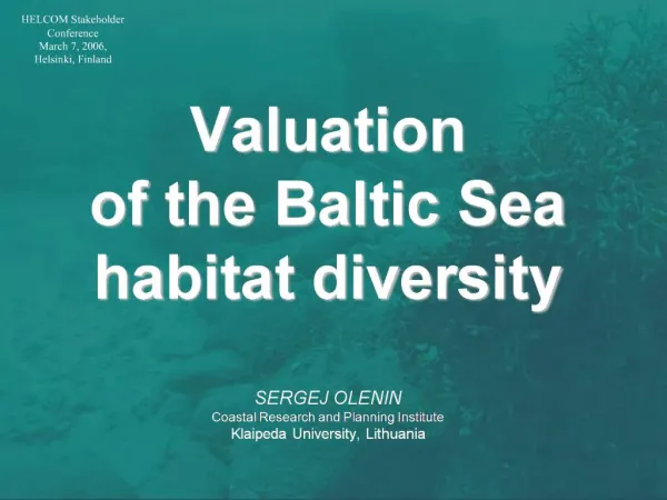
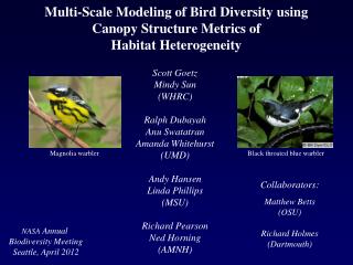
steadily goes down, probably due to the high Biswas and Paul (2012). Temperature, pH, salinity, peak in diatoms and filamentous cyanobacterial silicate of both water and sediment showed populations that increased in biomass and positively correlated with fungal diversity and occasional high values of nutrients, during the reactive silicate, inorganic phosphate, dissolved O2 seasons at salinities of 2-10%. Lowest oxygen level, phosphate, nitrogen, total organic carbon concentrations were detected in summer this showed a negative correlation. The data proposed situation probably occurs because respiration the overall maximum and minimum averages exceeds primary productivity in extremely strong among the stations was shown in Table 1. brines (Sammy (1983)). Phosphate level gradually decreases from station 1 to 3 due to Both Pielou’s and Shannon indices were highest the changes in the levels of phosphorous in at Marakkanamin station-III (5.151 and 9.319) sediment can be linked with the influx of (Table 2). Species richness and diversity of fungi phosphorous from upstream region and also its in all the sampling stations during the study regeneration into the overlying water column period was in conformity with the diversity under suitable conditions and the production of studies of Maria and Sridhar (2002). The K- H2S by anaerobic microbes enhances the dominance curves showed the cumulative eutrophication of phosphorus. dominance of species in rank order for each seasons. The fungal diversity showed variation in In the present investigation, total nitrogen season as well as stations wise. The maximum concentration was recorded high in monsoon diversity and density were observed in station 3 season and also gradual increase from reservoir during summer seasons (crystallizer pond) of to crystallizer. This is due to freshwater inflow both Tuticorin and Marakkanam saltpans. The which brings in abundant N2 rich terrigenous curve for the summer season of 2011 was lying deposits and their subsequent settling in at the bottom indicating highest diversity (Fig. 2). sediments and also statically significant correlation between sediment nitrogen and silt. In Tuticorin saltpans, based on the combination of Recent studies revealed that some proteinaceous delta+ and lambda+ values the number of species material could be resistant to microbial in the entire stations showed 95% confident limit degradation and that part of nitrogen may be except reservoir during summer in the year of preserved as such in sedimentary environments 2012, premonsoon and monsoon showed the (Nguyen and Harvey, 2001). High organic carbon exception in the year of 2012 in crystallizer ponds. was recorded at station-2 during monsoon and Low fungal counts were recorded as 20 species low in summer this may be due to the terrestrial whereas, in crystallizer pond during postmonsoon run-off that results in high levels of organic and summer had richest diversity in both years matter and inorganic nutrients (Rasheed et al., which contains 90 fungal species. Simultaneously, 2001). Similar observation were made during In Marakkanam saltpans, number of species in summer due to the decrease of organic carbon, every stations showed 95% confident limit except total nitrogen and silicate content in Secovlje in crystallizer pond during pre-monsoon in the salt-pans located in Gulf of Trieste, Northern year of 2011 and 2012 which contains 10 of fungal Adriatic (Kovac, 2009). The general decrease in species. In case of reservoir and evaporator TN and TOC contents might reflect an overall ponds, summer season showed the exception in decrease in primary productivity as salinity the year of 2012 which contains 40 of fungal increased (Liu et al., 2004). Lowering range of species and above. In crystallizer pond, summer season had richest diversity in both years which phosphate content were moderate in terms of contains 60 of fungal species (Fig. 3). total organic carbon was observed in the study of 6 Rani and Kalaiselvam
Sediments harbour more fungal counts than in Basidiomycota, 2 species in 2 genera fit in to water samples. Density of halophilic fungi ranged Zygomycota and the remaining 63 species in 15 from 3.0×10³ to 5.4×10⁴ CFU/ml in water sample genera belongs to mitosporic fungi which included and 3.6×10³ to 5.7× 10⁴ CFU/g in sediment sample 56 species in 13 genera of Hypomycetes and 2 of both Tuticorin and Marakkanam salterns. In both species in 2 genera of Coelomycetes respectively. the saltpans, the higher fungal density was 22 species of fungi were common to both the recorded in station-III. Overall, maximum fungal stations of Tuticorin and Marakkanam saltpans. counts were observed in summer season (2011) 16 species of fungi present only in Marakkanam while minimum in monsoon season (2012). In and 12 species of fungi was recorded in Tuticorin Tuticorin saltpan, 80 species belongs to 32 genera saltpan (Table: 3 & Fig. 3). were recorded, In Marakkanam saltpan, 83 species belongs to 36 genera were recorded. The predominance of Aspergillus species such as A. niger, A. terreus, A. fumigatus, A. candidus, A. Altogether, 95 species of halophilic fungi from wentii, A. flavus, A. sydowii, A. versicolor, A. water and sediment samples belongs to 41 ochraceus, A.Tamarii and Penicillium chrysogenum genera was recorded in both saltpans. Of these, were the common isolates identified during this 26 species in 21 genera falls under Ascomycota survey. A. niger was ranked first among the and 4 species under 3 genera belongs to halophilic fungal. Table 1. Physico-chemical parameters of water and sediment samples. Parameters Water Rainfall Atmospheric temperature Temperature pH Salinity Dissolved Oxygen Inorganic phosphate Nitrate Reactive silicate Sediment Temperature pH Salinity Total Nitrogen (TN) Total Phosphate (TP) Total Organic Carbon (TOC) Tuticorin Saltpan Marakkanam Saltpan 2.13 to 124.1 mm 22.8 to 37.5ºC 22 to 34oC 7.23 to 7.7 21 to 395‰ 1.43 to 4.35 mg/l 2.62- 4.33 µM/l 1.2- 5.1 µM/l 34.8 to 59.82 µM/l 0.66 to 128.7 mm 22.5-37ºC 20 to 35.8oC 7.2 to 7.6 19 to 400‰ 1.28 to 4.92 mg/l 2.62- 5.55 µM/l 1.32- 5.19 µM/l 31.9 to 60.64 µM/l 21 to 34oC 7.2 to 7.65 24 to 400‰ 4.43 to 8.52 µg/g 0.95 to 1.782 µg/g 2.37 to 7.48 mg/g 20 to 35.6oC 7.1 to 7.59 20 to 410‰ 4.36 to 8.58 µg/g 0.951 to 2 µg/g 2.51 to 7.59 mg/g Diversity, this may be due to the ability of this Other species of Aspergillus in teleomorphs found fungus to grow in the absence of oxygen and the in haline environments include Eurotium genus. production of pigmented spores that are more Kis-Papo et al. (2001) reported that the most resistant than hyaline spores to extreme frequently isolated species of halotolerant were E. conditions (Domsch et al., 1993). Penicillium was amstelodami, E. chevalieri, E. echinulatum, also in fairly high proportion that can able to E. halophilicum, E. herbariorum, E. intermedium, dwell in low water potential and low to high E.repens, E.rubrum, E. Spiculosum and E. temperature. The active mycota were dominated umbrosum both from arid, saline soil and salt by these two genera because of their marshes, in Israel, Syria and Kuwait. Butinar et osmoregulative mechanism and hence species of al. (2005) reported that E. Rubrum were Aspergillus and Penicillium could be isolated even recorded in the salterns only occasionally at lower from the deep sea environment (Jones, 1988). salinities (5–15% NaCl) at Dead Sea, Adriatic and 7 Rani and Kalaiselvam
Eilat salterns. In this study, similar results were crystallizer ponds at maximum salinity range. The observed that Eurotium amstelodami occurred fungal membranes are more fluid over a wide commonly from moderate to high concentration ranges of NaCl concentrations which might be the of NaCl and in case of Eurotium rubrum showed possible reason that indicating high intrinsic salt their presence occasionally. Kis-Papo et al. stress tolerance. (2003) reported that in vitro studies showed that the spores and mycelium of these species are The highly halotolerant fungus Gymnascella able to survive long-term exposure in solutions marismortui can well adapted to extreme within a broad range of salt concentrations (0– hypersaline environments. It was recorded in 30%). Similarly, 75% of all Penicillium sp. could water of the Dead Sea (range of 600‰) and was tolerate 20% NaCl and more than half of these never recorded on other localities (Buchalo et al., survived 25% NaCl (Tresner and Hayes, 1971). 1998). Interestingly, in crystallizer pond at In this study, 13 species were identified and Marakkanam saltpan, the isolates of ascomycete showed there occurrence at the salinity range of Gymnascella marismortui, were isolated at the 19-400‰. Kogej et al. (2005) reported that salinity range of 331-400‰. It is the first record minimal addition of NaCl slow down the growth that this species were isolated in Marakkanam rate of these species. Redkar et al. (1998) stated saltpan area in this study. Grishkan et al. (2003) that Emericella nidulans can adapt to gradually reported that only species of Gymnascella higher concentrations of NaCl. Here, in this study, marismortui was shown to be an obligate Emericella nidulans showed their occurrence halophile, which grown in the presence of 0.5-2 M occasionally at high concentration of salinity in of NaCl or 10-30% in Dead Sea water. In this both brine and saline soil. Thamizhmani et al. study, W. sebi and W. Ichthyophaga were (2013) reported the presence of Emericella both isolated at high concentration of NaCl and they in Marakkanam saltpan and marine ecosystem in showed their presence very rarely. Zalar et al. east coast of Tamilnadu. At high salinities the (2005) reported that W. ichthyophaga, W. muriae black yeast Hortaea werneckii, showed their and W. sebi can adapt in low concentration of dominance in the hypersaline waters of the NaCl, 5% for W. Muriae and W. sebi as in case of salterns. In this present work, this species were W. ichthyophaga it was found to be 15%. They found tobe common in both evaporation and can also withstand at high NaCl concentrations. Table 2. Species richness, diversity and evenness of Halophilic fungi recovered from all stations of saltpans. MargalfSpecies richness(D’) TUTICORIN STATIONS (RESERVOIR-I, EVAPORATOR-II, CRYSTALLIZER-III) III I II III 5.45 6.209 8.403 5.21 7.89 5.286 7.878 9.201 0.8191 4.55 4.277 5.005 5.738 5.179 0.8239 0.7982 0.8023 0.8284 0.7869 0.8049 4.348 3.836 3.833 4.371 4.103 4.038 4.905 4.452 3.771 4.827 4.885 4.271 0.8347 0.7605 0.8061 4.366 4.226 4.552 4.230 4.113 4.511 5.521 6.167 8.129 5.185 4.16 4.241 4.714 3.999 4.017 4.632 4.907 6.368 7.791 4.397 6.157 7.443 0.8111 0.7722 0.8154 0.8072 0.7402 0.8125 4.124 3.828 3.809 4.032 3.75 3.82 4.892 4.432 4.189 4.539 4.231 3.687 3.73 4.076 3.742 3.678 4.974 4.223 4.13 4.622 5.093 3.539 0.8122 0.7443 0.7678 0.8012 0.7183 Shannon Wiener Diversity (H’) TUTICORIN Pielou’sEvenness (J’) TUTICORIN SEASONS MARAKKANAM MARAKKANAM MARAKKANAM I II III I II I II III I II III I II III Pos-11 Sum- 11 Pre-11 Mon-11 Pos-12 Sum-12 Pre-12 Mon-12 4.326 4.352 4.757 4.513 4.679 5.014 4.267 4.438 4.872 4.376 4.784 5.151 5.148 6.324 4.355 4.026 3.937 4.347 4.243 4.193 5.234 7.904 9.319 0.8129 0.7924 0.8054 0.8538 0.7988 0.8236 0.808 0.8353 0.828 0.8201 0.8462 0.839 0.7877 0.815 7.09 7.667 0.8204 0.7741 0.7803 0.8121 0.7288 0.7876 5.02 4.212 0.7977 0.7657 0.7841 0.7992 0.7254 0.7864 0.792 Periodicities of occurrences of halophilic Basipetospora, Polypaecilum, Scopulariopsis and mycoflora diversity were tabulated based on its Cladosporium could tolerate much saline frequency in various stations of Tuticorin and extremes. In the present investigation, true Marakkanam saltpans (Table 4). Hooley et al. obligate halophilic fungal species and black yeasts (2003) reported the halophilic and halotolerant like Basidiobolus haptosporus, Bipolaris spicifera, yeast belong to genus Wallemia and Botryophialophora marina, Candida sp., Debaryomyces, Pichia, Rhodotorula, Hortaea, Cryptococcus neoformans, Dabaryomyces 8 Rani and Kalaiselvam
hansenii, Dreschlera halodes, D. hawaiiensis, Stachybotrys sp., Stenella araguata, Eurotium rubrum, Exophiala xenobiotica, Trichophyton verrucosom, Trimmatostroma sp., Exserohilum rostratum, Paecilomyces sp., T. salinum, Wallemia sebi and Yarrowia lipolytica Phaeotheca fissurella, Scopulariopsis brevicaulis, were isolated that can tolerate upto Fig. 2. K-Dominance curve representing the fungal diversity in season wise in both Tuticorin and Marakkanam salterns. Fig. 3. Ellipse plot representing overall fungal diversity of both Tuticorin and Marakkanam salterns. 9 Rani and Kalaiselvam
Table 3. List of fungi from saltpans of Marakkanam and Tuticorin. S. No Fungi Halophilic Fungi present only Tuticorin Botryophialophora marina Dabaryomyces hansenii Basidiobolus haptosporus Cryptococcus neoformans Exophiala jeanselmei Exophiala xenobiotica Gymnascella marismortui Hyphospora agavaciensis Non-sporulating fungi-4 Penicillium polonicum Phaeotheca fissurella Scedosporium apiospermum Stachybotrys sp. Stenella araguata Trichophyton verrucosum Exserohilum rostratum Halophilic Fungi common to both Stations Aspergillus candidus Aspergillus flavus Aspergillus fumigates Aspergillus niger Aspergillus sydowii Aspergillus melleus Aspergillus nidulans Aspergillus ochraceus Aspergillus restrictus Aspergillus sulphurous Aspergillus tamarii Aspergillus terreus Aspergillus versicolor Aspergillus ustus Aspergillus wentii Cladosporium cladosporioides Curvularia lunata Penicillium chrysogenum Penicillium fellutanum Penicillium digitatum Penicillium expansum Penicillium oxalicum Marakkanam Alternaria alternata Aspergillus sp. 1. 2. 3. 4. 5. 6. 7. 8. 9. 10. 11. 12. 13. 14. 15. 16. Exserohilum rostratum Non-sporulating fungi-3 Paecilomyces sp. Phaeotheca triangularis Scopulariopsis brevicaulis Sterile Black mycelium 3 Syncephalastrum racemosum Trimmatostroma salinum Wallemia ichthyophaga Aureobasidium sp. 17. 18. 19. 20. 21. 22. the salinity range of 395-410‰ in both readily greater numbers in sediment, while the evaporator and crystallizer ponds throughout the facultative halophiles were found more in the season of salt production (summer) at Tuticorin water column (Oren, 2003). This can be also due and Marakkanam saltpan. In the extreme to a physiological stress to stenohaline algal hypersaline conditions of both ponds, the species resulting mortality during increased salt environment is too harsh and biodiversity is concentration. The accumulated dead algae consequently limited while many taxonomic contributed to the high organic content of the groups are absent, but obligate halophilic and soil. Thamizhmani et al. (2013) reported among halotolerant taxa persist and thrive. This the 46 species of fungi isolated, 35 species were confirmed result was supported by Rodriguez- isolated from sediment samples followed by Valera (1988). Melanized fungi, a selective water with 31 species. In this present survey, advantage over the mycoflora in saline Marakkanam showed the maximum fungal environments, representing 85-100% of the total diversity than Tuticorin, this may be due to isolated mycobiota from highly saline waters and comparatively longer concentration process of partially replaced by non-melanized fungi at salts in addition to spatial changes would have lowered salinities, being detected only caused the occurrence of lower species diversity occasionally with NaCl concentrations below 5%. and population density in the Tuticorin saltpans. Similar observations were reported by In the present investigation, sediment acts as a Rajalekshmi (2001) in Marakkanam and Tuticorin good anchor for the fungi to dwell than water. saltpans, Tamilnadu. Nearly 80 species of fungi isolated from the saline soil of both study area this may be due to the In this study, the fungal density showed massive precipitation of halite from the water column by progress in crystallizer and evaporator ponds evaporation and so the salinity ranges high in during summer in this survey (2011-2012). sediment than water this could be a possible Likewise, fungal diversity and density showed reasons for obligate halophiles to survive more huge diverse at increased salinity. The 10 Rani and Kalaiselvam
hypothetical reasons behind may be at high environmental heterogeneity with a steep salinity salinities, often high densities of phototrophic gradient. In the summer seasons, increased brine microorganisms, planktonic as well as benthic, temperature and longer day length, both due to makes the salterns excellent living biological increased solar radiation, apparently promote system of primary production and other microbial biological productivity in solar salt ponds. While processes. In evaporator ponds, most of the the unadapted species gradually decreases while primary production occurs in benthic microbial increasing of salinity, thus the sole nutrients may mats seal ponds against water leakage and comes from the dead and decaying organic infiltration, permanently remove excess matter which helps the fungi to thrive well in such quantities of nitrogen and phosphate from the extreme hypersaline environments. Pedros-Alio overlying water and maintain desired thicknesses (2004) concluded that these ponds provide a in all ponds (Davis, 2000) and dominated by diversity of environments where different conditions different types of unicellular and filamentous of salinity, pH, temperature, light intensity, oxygen cyanobacteria sometimes in association with and nutrient concentrations are found, allowing the diatoms. Diatom diversity decreased noticeably study of different microbial communities. with increasing salinity, the number of benthic diatom taxa was much higher at lower salinities The findings showed the number of obligate (salinity 58–114g/l; 12 to 18 taxa) than in high halophilic fungi isolated in the present work salinities (salinity 157-206g/l; 1-5 taxa) in the seems to be higher as compared to that isolated salterns of Eilat, Israel (Oren et al., 2009). The on media without high salt concentrations. planktonic and benthic communities of marine Buchalo et al. (1998) stated that obligate organisms (e.g. bacteria, algae, copepods, halophilic fungi did not grow in freshwater molluscs, worms) that develop along with the medium and showed increased growth with increasing salinity gradient in the evaporating increasing salinity at 35ºC and the ionic ponds and crystallizers of saltworks create a composition of the medium had little effect on biological system that can either help nor harm growth. During monsoon season, microbial the salt production (Davis, 1993). growth was observed only in the salinity range of 19-35‰. However, during the entire study In crystallizer ponds, the unicellular green algae period it showed the occurrence of common Dunaliella are the sole primary producer that marine forms in the saltpans due to the inflow of lives in association with dense communities of marine water mixed with rain water and from halophilic archaea as well as extremely halophilic post monsoon onwards flourishing growth on the bacteria that color the brines red. Costa et al. all salinity ranges were observed. Extensive (1996) stated that solar salterns are not just salt studies were required for exploiting and production plants, they also function as acknowledge the fungi from solar salterns integrated saline wetlands of a unique coastal habitats especially in India. aquatic ecosystem that combines considerable Table 4. Periodicity of occurrence of halophilic mycoflora in various stations of Tuticorin and Marakkanam saltpans. FUNGI TUTICORIN S-2 (EVAPORATOR) W MARAKKANAM S-2 (EVAPORATOR) W S-1 S-3 S-1 S-3 (RESERVOIR) W (CRYSTALLIZER) W (RESERVOIR) W (CRYSTALLIZER) W S S S ASCOMYCOTA S S S - - S S S Bipolarisspicifera Botryophialophora marina Candida albicans Candida sp. Chaetomiumglobosum - - - - - - - - - - - - - - - - - - - - - - - S O - - - - - - - - - - - - S - O - - S - S - - S S S S - S S - 11 Rani and Kalaiselvam
FUNGI TUTICORIN S-2 (EVAPORATOR) W MARAKKANAM S-2 (EVAPORATOR) W S-1 S-3 S-1 S-3 (RESERVOIR) W (CRYSTALLIZER) W (RESERVOIR) W (CRYSTALLIZER) W S S S ASCOMYCOTA S - - O O - - - - C - - S - S - - S S S S S S S Chrysosporiumtropicum Dabaryomyceshansenii Eurotiumamstelodami Emericellanidulans Eurotiumrubrum Exophialajeanselmei Exophialaxenobiotica Exserohilumrostratum Gymnascellamarismortui Hortaeawerneckii Hyphosporaagavaciensis Phaeothecafissurella Phaeothecatriangularis Scedosporiumapiospermum Saccharomyces cerevisiae Stenellaaraguata Trichophytonverrucosum Trimmatostromasalinum Trimmatostromasp. Ulocladiumchartarum Yarrowialipolytica BASIDIOMYCOTA Cryptococcus neoformans Rhodosporiumsphaerocarpum Wallemiaichthyophaga Wallemiasebi MITOSPORIC FUNGI (HYPOMYCETES) Alternariaalternata Alternariasp. Aspergilluscandidus Aspergillusflavus Aspergillusfumigatus Aspergillusniger Aspergillussydowii Aspergillusflavipes Aspergillusglaucus Aspergillusjaponicus Aspergillusmelleus Aspergillusnidulans Aspergillusochraceus Aspergillusoryzae Aspergilluspenicilloides Aspergillusrestrictus Aspergillussclerotium Aspergillussp. Aspergillussulphureus Aspergillustamarii Aspergillusterreus Aspergillus unguis Aspergillusversicolor Aspergilluswentii Aspergillusustus Aureobasidiumpullulans Aureobasidiumsp. Cladosporiumcarrionii Cladosporiumcladosporioides Cladosporiumherbarum Cladosporiumsphaerospermum Curvularialunata Curvulariasp. Dreschlerahalodes Dreschlerabiseptata Dreschlerahawaiiensis Fusariumsolani Fusariumverticilliodes Nigrosporasphaerica Paecilomycessp. Penicilliumbrevicompactum Penicilliumchrysogenum Penicilliumfellutanum Penicilliumcrustosum Penicilliumcyclopium Penicilliumdigitatum Penicilliumexpansum Penicilliummarneffei Penicilliumoxalicum Penicilliumpolonicum Penicilliumsp. Penicilliumvariabile Penicillumcitrinum Scopulariopsisbrevicaulis Stachybotrys sp. Trichodermapiluliferum - - - - - - - - - - - - - - - - - - - - - - - - - - - - - - - - - - - - - - - - - - - - C S - - - - - S - - S - - - - - - S - S - C S - - - - - C - - S - - - - - - - - - S C - - - - S - C - - - - - - - S - S S - - - - - - - - - - - - - - - - - - - - - - - - - - - - - - - - - - - - - - - - - - S - - - - - - - - S S - - - S - - - - S - S - - S - - - - - C O - - - S - - - - - - S - C - O S S - S C S S - S S S S - S S S S - C O O S S - - C - - - - S - - - - C O - - - - - - - - - S S - - S S - - - - - - - S S - - - - - - - - - - - - - - - - S S - S - S - S - - - O MC MC C O O O - - MC C - C O - O O MC - C C C - - - C - - C - - - - - - - - O MC C O O MC MC - MC - - S S - - - - - O O MC MC C O O O S S MC O - C - - - C MC - C C C - - - C - - C - - - - C O - - C MC C O O MC MC O MC - - S S - - - - S S S MC MC C - - - - S MC S S C - - - O MC O MC C C O O S MC - - O - - - - - - S - - MC MC O - MC MC - C - - - - - - - - S S O MC MC C - O - O S MC S S C S - O O MC O MC C C O O C MC S - O O - - - - - S - O MC C O O MC MC - MC - - - - - - S - O - C MC MC MC S - - O - C - O C - - - O MC O C C C O C S MC - - S O O O S - - - - - MC MC - - MC MC - MC - - - - S - - - O S C MC MC C - - - - S C - O C - - S O MC O C C S O - - C - C S O - O S - - S S - MC C - - MC MC - C - O - - - - - - - S O MC MC C O - - O - MC S - C S - S O MC - C MC C - - - C - - C - - - - S O - - - MC C O - MC MC S MC - - - S - - - - - O O MC MC C O O O O O MC O - C O - O C MC - C MC C - - - C - - C - - - - C O - - O MC C O O MC MC O MC - - S O - - - S S O C MC MC MC O O - - O MC S S C - S O O MC O MC C C O - S MC S C O - - S - - O - - C MC MC O C MC MC S MC S O - O - - S S O S O MC MC C - O - O O MC S S C S S O O MC O MC C C O - S MC S C O - - S - O O S - C MC MC O C MC MC S C - S - O - - S S O - C MC MC MC O - - - O C - O C - - - O MC O C O S C - - MC - C O - - - S - - S - C MC MC - - MC MC - MC O O - - - S - - O S C MC MC C O - - O O C - O C - - S O MC O C O C C - S MC S C O O O O S - - S - C MC MC - C MC MC - C - S - - - - S 12 Rani and Kalaiselvam
FUNGI TUTICORIN S-2 (EVAPORATOR) W MARAKKANAM S-2 (EVAPORATOR) W S-1 S-3 S-1 S-3 (RESERVOIR) W (CRYSTALLIZER) W (RESERVOIR) W (CRYSTALLIZER) W S S S ASCOMYCOTA S S S MITOSPORIC FUNGI (COELOMYCETES) Non-sporulating fungi-1 Non-sporulating fungi-2 Non-sporulating fungi-3 Non-sporulating fungi-4 Sterile Black mycelium-1 Sterile Black mycelium-2 Sterile Black mycelium-3 ZYGOMYCOTA Basidiobolushaptosporus Syncephalastrumracemosum C - S - - - - - C - - - - S - - - - - S - - - - - - S - - - - - O - - - - - - - S - - - - S - - - C C - - - S - C - - - S - - C O - - - - - - - - - - - - - O - - S - - - - - O - - - - - - - - - - - - - - - - - - S - 13 Rani and Kalaiselvam
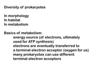
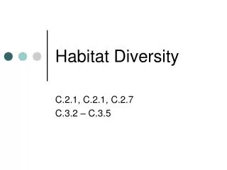
Fig. 3. Microscopic observation of halophilic fungi. References Buchalo AS, Nevo E, Wasser SP, Oren A, Anbazhagan P. 1988. Hydrobiological and Molitoris HP. 1998. Fungal life in the extremely benthic ecology of Kodiakkarai coastal sanctuary hyper saline water of the Dead Sea: First records. (Southeast coast of India). Ph.D Thesis, Proceedings of Royal Society of London 265, Annamalai University, India 208. 1461-1465. Baati H, Guermazi S, Amdouni R, Gharsallah Butinar L, Santos S, Spencer-Martins I, Oren N, Sghir A, Ammar E. 2008. Prokaryotic A, Gunde Cimerman N. 2005a. Yeast diversity diversity of a Tunisian multipond solar saltern. in hyper saline habitats. FEMS Microbiology Extremophiles12, 505-518. Letters 244, 229-234. Biswas A, Paul AK. 2012. Physico-chemical Butinar L, Zalar P, Frisvad JC, Gunde analysis of saline soils of solar salterns and Cimerman N. 2005. The genus Eurotium - isolation of moderately halophilic bacteria for poly members of indigenous fungal community in (3-hydroxybutyric acid) production. International hyper saline waters of salterns. FEMS Research Journal of Microbiology2, 227-236. Microbiology Ecology 51, 155-166. 14 Rani and Kalaiselvam
Costa LT, Farinha JC, Hecker N, Tomas-Vives Gunde Cimerman N, Zalar P, Petrovic U, Turk P. 1996. Mediterranean wetland inventory:A M, Kogej T, de Hoog GS, Plemenitas A. 2004. reference manual. MedWet/Instit. Conserv. Natur./ Fungi in the Salterns. In: Halophilic Wetlands Int. Publications. Lisboa 1, 1-109. microorganisms, Ventosa, A., (Eds.), Springer- Verlag, Heidelberg 103-113. Davis JS. 1993. Biological management for Hooley P, Fincham DA, Whitehead MP, problem solving and biological concepts for a new Clipson NJW. 2003. Fungal osmotolerance. generation of solar salt works, Seventh Advance in Applied Microbiology 53, 177-211. Symposium on Salt 1, 611-616. Javor BJ. 2002. Industrial microbiology of solar Davis JS. 2000. Function and management of salt production. Journal of Indian Microbiology the biological system for seasonal solar salt work. and Biotechnology 28, 42-47. Global Nest. International Journal 2, 217-226. Jones EBG. 1988. Do fungi occurring the sea?. Domsch KH, Gams W, Anderson TH. 1993. Mycologist 2, 150-157. Compendium of Soil Fungi, Vol 1. IHW-Verlag, Kamat TK, Kerkar S. 2011. Pharmaceutical Eching. potentials of bacteria from saltpans of Goa, India. Dundas IE, Halvorson HO. 1966. Arginine International Journal of Pharmaceutical metabolism in Halobacteriumsalinarium, an Applications 2, 150-154. obligately halophilic bacterium. Journal of Kirk PM, Cannon PF, David JC, Stalpers JA. Bacteriology 91, 2001. Ainsworth and Bisby’s Dictionary of the 113-119. Fungi, IX (Eds.), CAB International, Oxon UK. Ehrlich DIA. 1987. The Effect of salinity and Kis-Papo T, Grishkan I, Oren A, Wasser SP, temperature gradients on the distribution of littoral Nevo E. 2001. Spatiotemporal diversity of microalgae in experimental solar ponds, Dead Sea, filamentous fungi in the hyper saline Dead Sea. Israel. P.S.Z.N.I: MarineEcology 8, 193-205. Mycological Research 105, 749-756. Ellis MB. 1971. Dematiaceous Hyphomycetes. Kis-Papo T, Oren A, Wasser SP, Nevo E. The Common wealth Mycological Institute, Kew, 2003. Survival of filamentous fungi in hypersaline Surrey, England. Dead Sea water.MicrobialEcology 45, 183-190. Femitha RD, Vaithyanathan C. 2012. Physico- Kogej T, Ramos J, Plemenitas A, Gunde chemical parameters of the various stages in Cimerman N. 2005. The halophilic fungus different Salt-pans of Tuticorin district. Journal Hortaea werneckii and the halotolerant fungus of Chemical and Pharmaceutical Research 4, Aureobasidium pullulans maintain low 4167- 4173. intracellular cation concentrations in hypersaline Griffith DH. 1994. Fungal Physiology, John Wiley environments. Applied Environmental and and Sons, New York 472. Microbiology 71, 6600-6605. Grishkan I, Nevo E, Wasser SP. 2003. Soil Kovac N. 2009. Chemical characterization of micromycete diversity in the hypersaline Dead stromatolitic “Petola” layer (Secovlje salt-pans, Sea coastal area (Israel). Mycology Slovenia) using FT-IR spectroscopy. ANNALES. Programme 2, 19-28. Series Historia Naturalis 19, 95-102. 15 Rani and Kalaiselvam
Lambshead JD, Platth M, Shawk M. 1983. The Nguyen RT, Harvey HR. 2001. Preservation of detection of differerences among assemblages of protein in marine systems: Hydrophobic and marine species based on an assessment of other non-covalent association as major dominace and diversity. Journal of Natural stabilizing forces. Geochimica et Cosmochimica History 17, 859-874. Acta 65, 467-1480. Litchfield CD. 1991. Red-the magic color for Nikolaou E, Agrafioti I, Stumpf M, Quinn J, solar salt production. In: Das Salz in der Rechts- Stansfield I, Brown AJP. 2009. Phylogenetic und Handelsgeschichte, Hocquet J.C. and R. diversity of stress signalling pathways in fungi, Palme, (Eds.), Berenkamp, Schwaz 403-412. BMC Evolutionary Biology 9, 44-62. Liu X, Cai K, Yu S. 2004. Geochemical Oren A, Bina D, Ionescu D, Prasil O, Rehakova simulation of the formation of brine and salt K, Schumann R, Sorensen K, Warkentin M, minerals based on Pitzer model in Caka Salt Lake. Woelfel J, Zapomelova E. 2009. Saltern Science47, 720-726. evaporation ponds as model systems for the study of microbial processes under hypersaline conditions- Madkour F, Gaballah MM. 2012. Phytoplankton An interdisciplinary study of the salterns of Eilat, assemblage of a solar saltern in Port Fouad, Israel. Proceedings of the 2nd International Egypt.Oceanologia 54, 687-700. Conference on the Ecological Importance of Solar Saltworks.Merida, Yucatan Mexico. Manikandan M, Kannan VR, Pasic L. 2009. Diversity of microorganisms in solar salterns of Oren A. 2002. Diversity of halophilic Tamil Nadu, India. World Journal of Microbiology microorganisms: Environments, phylogeny, and Biotechnology 25, 1007-1017. physiology and applications. Journal of Industrial Microbiology and Biotechnology 28, 56-63. Maria GL, Sridhar KR. 2002. Richness and diversity of filamentous fungi on woody litter of Oren A. 2003. Physical and chemical limnology mangroves along the west coast of India. Current of the Dead Sea. In: Fungal life in the Dead Sea, Science 83, 1573-1580. Nevo, E., A. Oren and S.P. Wasser, (Eds.), Gantner Verlag, Ruggel 45-67. Nazareth S, Gonsalves V, Nayak S. 2012. A first record of obligate halophilic Aspergilli from the Dead Pedros-Alio C, Calderon-Paz JI, MacLean Sea. Indian Journal ofMicrobiology 52, 22-27. ML, Medina G, Marrase C, Gasol JM, Guixa- Boixereu N. 2000. The microbial food web NealsonKH, Stahl S. 1997. Microorganisms and along salinity gradients. FEMSMicrobiology biogeochemical cycles: What can we learn from Ecology 32, 143-155. layered microbial communities?. In: Geomicrobiology, Banfield, J. and K.H. Nealson, Pedros-Alio C. 2004. Trophic ecology of solar (Eds.), Mineralogical Society of America, salterns. In: Halophilic Microorganisms, Ventosa, Washington, DC 1-34. A., (Eds.), Springer-Verlag Heidelberg 33-48. Nedumaran T, Perumal P. 2012. Biodiversity of cyanobacteria from Uppanar estuary, Southeast Petrovic A. 1998. Diatoms from the saltern of Coast of India. Emirates Journal Food and Ston (Croatia). Rapport Commission International Agriculture 24, 248-254. Mer Mediterranean35,574-575. 16 Rani and Kalaiselvam
Pielou EC. 1966. The measurement of diversity Shannon CE, Wiener W. 1949. The in different types of biological collections. Journal mathematical theory of communication. Univ. of. of Theoretical Biology 13,144. Ilinois Press. Urbana. Radhika D, Veerabahu C, Nagarajan J. 2011. Strickland JDH, Parsons TR. 1972. A practical Distribution of Phytoplankton and Artemia in the handbook of seawater analysis. Fisheries Solar Salterns at Tuticorin. Current World Research Board. Canada Bulletin 167, 310. Environment 6,233-239. RajalekshmiG. 2001. Distribution of fungi in the Subramanian B, Mahadevan M. 1999. Seasonal and diurnal variation of Hydrobiological characters hypersaline (Solar salterns) environs of Tamil Nadu, M.Sc., dissertation, CAS in Marine Biology, of coastal waters of Chennai Bay of Bengal. Indian Annamalai University, India 18. Journal of Marine Sciences 28, 429-433. Rasheed MA, Rodes CA, Thomas R. 2001. Subramanian SK, Kannan L. 1998. Poprt of mackey seagrass, algae and macro- Environmental parameters of the Indian marine invertebrate communication. CRC Reef Research biosphere reserve off Tuticorin in the Gulf of Centre technical report No. 43, Townsville. Mannar. Seaweed Research Utilization 20, 85-90. Redkar RJ, Herzog RW, Singh NK. 1998. Thamizhmani R, Vimal Raj R, Sivakumar T. Transcriptional activation of the Aspergilllus 2013. Abundance and diversity of fungi in salt pan nidulans gpd, A promoter by osmotic signals. Appl. Environ. Microbiol 64, 2229-2231. and marine ecosystem of the east coast of Tamil Rodriguez-Valera F. 1988. Characteristics and Nadu, India. International Journal of Current Microbiology and Applied Sciences 2, 67-75. microbial ecology of hyper saline environments. In: Halophilic bacteria, Rodriguez-Valera, F. and Tresner HD, Hayes JA. 1971. Sodium chloride F.L. Boca Raton, (Eds.), CRC Press 3-30. tolerance of terrestrial fungi. Applied Microbiology Sammy N. 1983. Biological systems in North- 22, 210-213. Western Australian solar salt fields. In: Sixth Turk M, Montiel V, Zigon D, Plemenitas A, symposium on salt, Schreiber, B.C and H.L. Harner Ramos J. 2007. Plasma membrane composition of (Eds.), The salt Institute, Toronto 1, 207-215. Debaryomyceshansenii adapt to changes in pH and Satyanarayana T, Raghukumar C, Shivaji S. external salinity. Microbiology 153, 3586-3592. 2005. Extremophilic microbes: Diversity and perspectives, Current Science 89, 78-90. Zalar P, de Hoog GS, Schroers HF, Frank JM, Selbmann L, de Hoog GS, Mazzaglia A, Gunde Cimerman N. 2005. Taxonomy and phylogeny of the xerophilic genus Wallemia Friedmann EI, Onofri S. 2005. Fungi at the (Wallemiomycetes and Wallemiales, Nov.). edge of life: Cryptoendolithic black fungi from AntonievanLeeuwenhoek 87, 311-328. Antarctic deserts. StudiesinMycology 51, 1-32. 17 Rani and Kalaiselvam