Download
1 / 6
0 likes | 11 Views
We characterized through morphological features and some biochemical tests bacterial isolates from the skin of three endemic frog species: Kalophyrnus sinensis (Peters, 1867), Limnonectes magnus (Stejneger, 1910), and Megophrys stejnegeri (Taylor, 1920) from Mt. Andapon Barangay Campawan, in Baganga, Davao Oriental, Philippines. The bacterial isolates were acquired through skin swabs from five representative adult individuals per species, grown in select solid media, and subjected to various standard biochemical tests. Nine bacterial isolates were obtained:

E N D
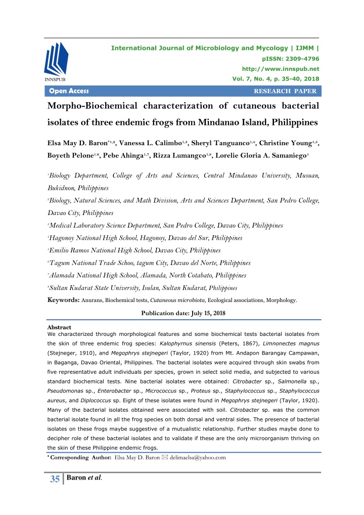
International Journal of Microbiology and Mycology | IJMM | pISSN: 2309-4796 http://www.innspub.net Vol. 7, No. 4, p. 35-40, 2018 Open Access RESEARCH PAPER Morpho-Biochemical characterization of cutaneous bacterial isolates of three endemic frogs from Mindanao Island, Philippines Elsa May D. Baron*1,2, Vanessa L. Calimbo1,3, Sheryl Tanguanco1,4, Christine Young1,5, Boyeth Pelone1.6, Pebe Ahinga1,7, Rizza Lumangco1,8, Lorelie Gloria A. Samaniego1 1Biology Department, College of Arts and Sciences, Central Mindanao University, Musuan, Bukidnon, Philippines 2Biology, Natural Sciences, and Math Division, Arts and Sciences Department, San Pedro College, Davao City, Philippines 3Medical Laboratory Science Department, San Pedro College, Davao City, Philippines 4Hagonoy National High School, Hagonoy, Davao del Sur, Philippines 5Emilio Ramos National High School, Davao City, Philippines 6Tagum National Trade Schoo, tagum City, Davao del Norte, Philippines 7Alamada National High School, Alamada, North Cotabato, Philippines 8Sultan Kudarat State University, Isulan, Sultan Kudarat, Philippines Keywords: Anurans, Biochemical tests, Cutaneous microbiota, Ecological associations, Morphology. Publication date: July 15, 2018 Abstract We characterized through morphological features and some biochemical tests bacterial isolates from the skin of three endemic frog species: Kalophyrnus sinensis (Peters, 1867), Limnonectes magnus (Stejneger, 1910), and Megophrys stejnegeri (Taylor, 1920) from Mt. Andapon Barangay Campawan, in Baganga, Davao Oriental, Philippines. The bacterial isolates were acquired through skin swabs from five representative adult individuals per species, grown in select solid media, and subjected to various standard biochemical tests. Nine bacterial isolates were obtained: Citrobacter sp., Salmonella sp., Pseudomonas sp., Enterobacter sp., Micrococcus sp., Proteus sp., Staphylococcus sp., Staphylococcus aureus, and Diplococcus sp. Eight of these isolates were found in Megophrys stejnegeri (Taylor, 1920). Many of the bacterial isolates obtained were associated with soil. Citrobacter sp. was the common bacterial isolate found in all the frog species on both dorsal and ventral sides. The presence of bacterial isolates on these frogs maybe suggestive of a mutualistic relationship. Further studies maybe done to decipher role of these bacterial isolates and to validate if these are the only microorganism thriving on the skin of these Philippine endemic frogs. * Corresponding Author: Elsa May D. Baron delimaelsa@yahoo.com 35 Baron et al.
Introduction the skin of three Philippine endemic frogs The Philippine tropical forest is home to several sampled from Mindanao and additional endemic frog species with increasing frequency of information on cutaneous bacterial isolates from new species discovered almost every year (Brown, frogs of the Philippines De Layola, Lorenzo, and Diesmos, 2015; Mcleod et al, 2011; Supsup, Guinto, Redoblado, and Gomez, Materials and methods 2017). Majority of the studies conducted are Isolation of cutaneous bacteria inventories focused on identifying what species are Five individuals each of Kalophrynus sinensis, present in different habitat types and deciphering Limnonectes magnus, and Megophrys stejnegeri elevational distribution (Sanguilla et al, 2016; were collected from a forest interior of Mt. Diesmos, Brown, and Gee, 2003; Siler et al, Andapon, Sitio Casunugan, Barangay Campawan, 2012). Data on ecology on these endemic frog Baganga, Davao Oriental, Mindanao, Philippines. species remains depauparate and limited reports Andapon Mountains form part of the Mt. Mayo- are published pertinent to microbial species Tagub Kampalili Complex, one of the identified assciated with frog species (Diesmos, Brown, and major Key Biodiversity Area of the country. Adult Gee, 2003; Ates and Delima, 2008; Warquez, frog individuals were handpicked from the site Mondejar, and Demayo, 2013; Dacalus et al, and transported through native bamboo basket to 2017). the site were swabbing was done. The handlers Ecological wore nitrile gloves to prevent transfer of interactions such as microbial microbes from the skin of the handlers to the associations in frogs were previously reported frogs’ skin. Prior to swabbing, frog individuals (Conlon, 2011;, McKenzie et al, 2012). Microbial were washed with sterile water twice for about 20 symbionts especially those found in the seconds on both dorsal and ventral sides to cutaneous region has been identified to have remove transient bacterial species (Woodhams et substantial significant influence in the capability al, 2007). Fresh nitrile gloves were used to handle of frogs to combat pathogens. Skin microbiome the frogs prior to swabbing. Each frog individual play as positive inducer for release of was swabbed twice: dorsal and ventral sides, antimicrobial peptides in frog’s skin of some using separate fresh, sterile swabs, 20 times each anuran species (Mangoni et al, 2001; Becker et side in order to get ample inoculum from the tip of al, 2014). Moreover species-specific resistance to the snout to the webbings of the hindfoot’s digits. pathogens such as fungi maybe influenced by Swabs for each side and individuals per species host-specific bacteria (Mangoni et al, 2001). were directly inoculated in separate tubes in Bacterial taxa in frog’s skin may also act as freshly prepared nutrient broth in the field barrier against pathogens, thereby forming a (Rahman, Somsiri, Tajima, and Ezura, 2004). Frog potential safetynet for the frog host (Sanguilla et individuals were released immediately after al, 2016). swabbing. Tubes were incubated at 250C for 24 Despite increasing number of literature reports hours. Subsequently, one ml inoculum from each turbid tube were diluted serially, three-fold and on the seemingly important role of bacterial poured on freshly prepared Trypticase soy agar, symbionts of the frog’s skin, we found only one nutrient agar, Mannitol-salt agar, and MacConkey report on bacterial taxa associated with Philippine agar plates. Pure cultures of each of the colonies frog species (Eda, Diesmos, Vredenburg, and were obtained and utilized for morphological and Chan, 2015). Thus this study provides baseline biochemical characterizations. information on bacterial isolates associated with 36 Baron et al.
Morpho-Biochemical characterization of bacterial these frogs are ground dwelling species isolates (International Union for the Conservation of Bacterial isolates obtained from plates with solid Nature, 2004; Inger, 1954). medium were initially characterized based on The variation in the number of isolates obtained coloration of the colony/colony forming unit, per frog species maybe attributed to the surface shape of margins of the colony, pattern of growth area available for the bacterial isolates to (after 24 hours), capacity to ferment lactose, and colonize. Among the three species, K. sinensis Gram staining reactions. Moreover biochemical has the least wide dorsum and ventral surface tests were performed for each isolate that have (International Union for the Conservation of grown in McConkey and Mannitol-Salt agar Nature, 2004; Inger, 1954) area which could plates. The biochemical tests performed included: explain why it has the least number of bacterial triple sugar iron agar (TSI), Lysine iron agar isolates. Although L. magnus, attributed to its (LSI), urease, citrase, coagulase, sulfide and stocky body (International Union for the indole formation and motility following standard Conservation of Nature, 2004; Siler McVay, procedures. Diesmos, and Brown, 2009), provides a wider Results and discussion area for bacterial species colonization, the Bacterial isolates obtained number of isolates obtained was way less compared Nine bacterial isolates were obtained and to that of M. stejnegeri. One reason for such maybe morpho-biochemically characterized from the the presence of pronounced ridges on the dorsum swabs of Kalophrynus sinensis (Peters, 1867), of L. magnus compared to the lesser ridges and Limnonectes magnus (Stejneger, 1910), and tubercles of M. stejengeri dorsum (International Megophrys stejnegeri (Taylor, 1920) (Table 1). Union for the Conservation of Nature, 2004; Siler Eighty eight percent (88%) of the bacterial McVay, Diesmos, and Brown, 2009). The presence isolates were obtained from M. stejnegeri while of the ridges may have influenced the inoculum the least number of bacterial isolates was obtained through swabbing as ridges may limit obtained from K. sinensis. Majority of these areas that can be reached by the tip of the sterile bacterial isolates are reported to be thriving in swab and such could possibly hamper access to soil (Tortora, Funke, and Case, 2013). The other bacterial isolates residing on areas proximal to presence then of these bacterial isolates in the the ridges. skin of the frogs examined is expected since Table 1. Morpho-biochemical characters of the bacterial isolates from three endemic frogs sampled from Mindanao, Philippines. Putative Id of bacterial isolate Morphological characteristics Biochemical Tests Results Frog Species Color Shape GSR LF TSI LIA Urease Citrase Coagulase Sulfide Indole Bacilli Mucoid Motile Citrobacter sp. (D, V) NLF, LF A, A +, + -, + White - + + + +, - Salmonella sp. (D) Pseudomonas sp. (V) Citrobacter sp. (D, V) Salmonella sp. Bacilli White K, A +, - K, K +,- A, A +,+ -,+ +, - - NLF + + +, - + mucoid colorless - NLF -,+ + + - - L. magnus White Bacilli - LF + + + +. - White Bacilli - NLF K, A -, + + - - 37 Baron et al.
(D) Pseudomonas sp. (V) Enterobacter sp. (V) Micrococcus sp. (D) Citrobacter sp. (D, V) +, - + K, K -,- A, A +,- colorless Bacilli - NLF -,+ + + - - Mucoid -, + colorless Bacilli - LF + + - - White Cocci + - Baccili Mucoid LF, LF A, A +, + K,A K,K -, + -, + Pink - + + + + Proteus sp. (D,V) +, - Colorless Mucoid - NLF + + + + M. stejnegeri Staphylococcus sp. (D) Enterobacter sp. (D) Staphylococcus aureus (D) Cocci in cluster White + Bacilli Mucoid Cocci in cluster K, A +, - -, + Pink - LF + + - + White + Pseudomonas sp. (D) Colorless Bacilli K, K +, - -, + - NLF + + - + Mucoid M. stejnegeri Diplococcus sp. (D) Cocci in pairs White + - Side obtained: D- dorsal side, V- ventral side; GSR- Gram Stain Reaction; LF-Lactose Fermentation: LF- lactose fermenter, NLF- Non-lactose fermente. Morpho-Biochemical results of the bacterial skin of some Philippine frogs in Luzon (Eda, isolates Diesmos, Vredenburg, and Chan, 2015). These Morphological assesment and biochemical test isolates were also found to be present in non- results revealed nine bacterial isolates from the Philippine frog species. Pseudomonas sp. was three frog species sampled (Table 1). Citrobacter documented in Atelopus spp. from Colombia sp. was the common isolate found in all frog (McKenzie et al, 2012; Harris et al, 2006). species swabbed which was obtained in both Citrobacter, Micrococcus, Enterobacter were also dorsal and ventral sides. Aside from an occasional earlier reported as inhabitants of the skin of pathogen, this bacterium can also be isolated amphibians (Carr et al, 1976; Hird et al, 1983). from soil (Tortora, Funke, and Case, 2013). Finding this bacterial taxon common in all frog The presence of bacterial isolates on frog’s skin samples is not surprising. The low number of is considered a mutualistic relationship. isolates that we obtained despite working with Bacterial species inhabiting the skin of various three varied frog species may suggest that frog frog species provide positive inducement for species harbor host-specific bacterial species release of antimicrobial peptides while others (McKenzie et al, 2012). Moreover, since the study block establishment of certain pathogens focused only on bacterial isolates that have (Mangoni et al, 2001; Becker et al, 2014). grown on the media used, other bacterial isolates However it is not yet fully established if all thriving on the frogs’ skin may have not been bacterial taxa residing in the skin of frogs accounted. accomplish both these functions. The bacteria Two of the bacterial isolates (Enterobacter sp., and seem to benefit from this relationship by the nourishment provided by the mucus secretions Pseudomonas sp.) reported in this study were of the frog’s skin (Lauer et al, 2007). previously accounted to be associated with the 38 Baron et al.
Conclusion Carr AH, Amborski RL, Culley DD, Amborski Nine bacterial isolates from the skin of three GF. 1976. Aerobic bacteria in the intestinal tracts endemic frog species: Kalophyrnus sinensis of bullfrogs (Rana catesbeiana) maintained at low (Peters, 1867), Limnonectes magnus (Stejneger, temperatures. Herpetologica 32, 239–244. 1910), and Megophrys stejnegeri (Taylor, 1920) Conlon J. 2011. The contribution of skin were obtained. Isolates which were characterized antimicrobial peptides to the system of innate thorugh morphological and results of some immunity in anurans. Cell Tssue Res 343, 201- biochemical tests incluuded Citrobacter sp., 212. Salmonella sp., Pseudomonas sp., Enterobacter sp., Micrococcus sp., Proteus sp., Staphylococcus Dacalus C, Calunsag A, Hoshino L, Peralta D, sp., Staphylococcus aureus, and Diplococcus sp. Baron E. 2017. Anuran assemblage on forest edges Eight of these isolates were found in Megophrys of Datu Salumay, Davao City, Philippines. University stejnegeri (Taylor, 1920). Citrobacter sp. was the of Mindanao International Multidisciplinary Research common bacterial isolate found in all the frog Journal. species on both dorsal and ventral sides. The https://doaj.org/article/38a3a4293c7247f1a8115b8 presence of bacterial isolates on these frogs acd90 2ce2. maybe suggestive of a mutualistic relationship, Diesmos though this requires further studies. A, Brown R, Gee G. 2003. Acknowledgment International Union for the Conservation of Preliminary Report on the Amphibians and The researchers are grateful to the logistical support generously provided by Dr. Ana Julia Reptiles of Balbalasang-Balbalan National Park in Enero, Dean of College and to Prof. Helen Ancla, Luzon Island, Philippines. Slyvatrop The Technical Head of laboratories of San Pedro College, Davao Journal of Philippine Ecosystems and Natural City. We also acknowledge the field assitance Resources13, 63-80. rendered by the locals of Mt. Andapon, Baganga, Eda A, Diesmos A, Vredenburg V, Chan M. Davao Oriental. 2015. Isolation, Screening, and Identification of References Frog Cutaneous Bacteria for Anti- Ates F, Delima EM. 2008. Assemblage and Batrachochytridium dendrobatidis Activity. microhabitats of anurans from Mt. Sinaka, Arakan, Interantional Journal of Bioengineering and Life Cotabato and Mt. Hamiguitan, Davao Oriental, Sciences 9(4). Mindanao Island, Philippines. Journal of Nature Flechas S, Sarmiento C, Cardenas M, Medina Studies 7(1), 101-107. Becker M, Zawacki C, Gratwicke B, and Bleden E, Restrepo S, Amezquita A. 2012. Surviving Chytridiomycosis: differential anti- L. 2014. The effect of captivity on the cutaneous Batrachochytridum dendrobatidis activity in bacterial community of the critically endgangered bacterial isolates from three lowland species of Panamian golden frog (Atelopus zeteki). Biological Atelopus. Plos One 7(9), 1-7. Conservation 176, 199-206. Harris R, James T, Lauer A, Simon M, Patel Brown R, De Layola L, Lorenzo A, Diesmos A. 2006. Amphibian pathogen M. 2015. A new species of limestone karst Batrychochytridium dendrobatidis is inhibited by inhabiting forest frog, genus Platymantis the cutaneous bacteria of amphibian species. (Amphibia: Anura: Ceratobatrachidae. Zootaxa EcoHealth 3, 53-56. 4048(2), 191-210. 39 Baron et al.
Hird DW, Diesch SL, McKinnell RG, Gorham Sanguilla M, Cobb K, Siler C, Diesmos A, E, Martin FB, Meadows CA, Gasiorowski M. Alcala A, Brown R. 2016. The amphibians and 1983. Enterobacteriaceae and Aeromonas- reptiles of Mindanao Island, southern Philippines, Hydrophila in Minnesota frogs and tadpoles (Rana II: the herpetofauna of northeast Mindanao and pipiens). Applied and Environmental adjacent islands. ZooKeys 642, 1-132. Microbiology 46(6), 1423-1425. Inger RF. 1954. Systematics and zoogeography Siler C, McVay J, Diesmos A, Brown R. 2009. A new species of fanged frog, genus Limnonectes, of Philippine amphibia. Fieldiana 33, 181-531. (Amphibia: Anura: Dicroglossidae) from Southeast International Union for the Conservation of Mindanao Island, Philippines. Herpetologica 65(1), 105-114. Nature, Conservation International and Nature Reserve. 2004. Global amphibian Siler C, Swab J, Oliveros C, Diesmos A, Averia assessment. 15 April 2018. L, Alcala A, Brown R. 2012. Amphibians and www.Globalamphibians.org. reptiles , Romblon Island Group, central Philippines: Lauer A, Simon MA, Banning JL, Andre ́ E, comprehensive herpetofaunal inventory. Checklist Duncan K, Harris R. 2007. Common cutaneous 8(3), 443-462. bacteria from the eastern red-backed salamander can inhibit pathogenic fungi. Copeia 3, 630–640. Supsup C, Guinto F, Redoblado R, Gomez R. Mangoni M, Miele R, Renda T, Barra D, 2017. Amphibians and reptiles from the Mt. Hamiguitan Range of eastern Mindanao, Simmaco M. 2001. The synthesis of Philippines: New distribution records. Checklist antimicorbial peptides in the skin of Rana 13(3), 2121. esculenta is stimulated by microorganisms. FASEB Journal 15(8), 1431-1432. Tortora G, Funke B, Case L. 2013. Microbiology McKenzie V, Bowers R, Fiere N, Knight R, an introduction 11th edition. Pearson, Boston USA. Pp. 975. Lauber C. 2012. Co-habiting amphibian species harbor unique skin bacterial communities in wild Warguez D, Mondejar E, Demayo C. 2013. Frogs populations. International Society for Microbial and their microhabitat preferences in the Ecology Journal 6, 588-596. agricultural and secondary forest areas in the Mcleod D, Siler C, Diesmos A, Diesmos M, vicinity of Mt. Kalatungan Mountain, Bukidnon, Garcia V, Arkonceo A, Balaquit K, Uy C, Philippines. International Research Journal of Villaseran M, Yarra E, Brown R. 2011. Biological Sciences 2(10), 51-63. Apmhibians and Reptiles of Luszon Island V: The Herpetofauna of Angat Dam Watershed, Bulacan Woodhams D, Vredenburg V, Simon M, Province, Luzon Island, Philippines. Asian Billheimer D, Shakhtour B, Shyr Y, Briggs C, Herpetological Research 2(4), 177-198. Smith L, Harri R. 2007. Symbiotic bacteria Rahman M, Somsiri T, Tajima K, Ezura Y. contribute to innate immune defenses of the threatened mountain yellow-legged frog, Rana 2004. Distribution of Aeromonas spp. muscosa. Biological Conservation 138, 390-398. Emphasizing on a newly identified species Aeromonas sp. T8 isolated from fish and aquatic animals in Southeast Asia. Pakistan Journal of Biological Sciences 7(2), 258-268. 40 Baron et al.