Download
1 / 33
370 likes | 477 Views
Discover the categorization and functions of spinal nerves and sensory receptors in the peripheral nervous system. Explore how proprioceptors and motor endings work, along with the organization of spinal nerves. Learn about nerve plexuses and their role in innervation, including the cervical plexus.

E N D
Peripheral Nervous Systemways to categorize: • Motor or sensory • General (widespread) or specialized (local) • Somatic (outer tube) or visceral (inner tube)
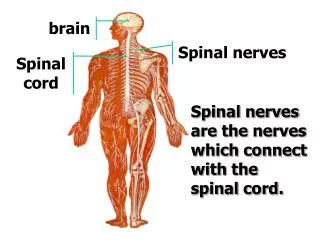
____Cranial nerves attach to brain ___Spinal nerves attach to spinal cord
Peripheral sensory receptors By location: • Exteroceptors • Sensitive to stimuli arising from outside body • Interoceptors • Or visceroreceptors, from internal viscera • Proprioceptors • Monitor degree of stretch in skeletal muscles, tendons, joints and ligaments
Sensory Receptors • Free nerve endings (pain and temp) • Merkel discs (light touch) • Root hair plexuses – entwine hair follicles (light touch) • Encapsulated Meissner’s corpuscles (light touch in hairless skin) • Ruffini’s corpusucles (deep pressure and stretch) • Pacinian corpuscles (deep pressure, vibration, visceral: pain, nausea, hunger, fullness)
Proprioceptors • Skeletal muscles, joints, tendons, ligaments • Degree of stretch, therefore information on body movement: • to cerebrum, • cerebellum and • spinal reflex arcs • Include: -Muscle spindles -Golgi tendon organs -Joint kinesthetic receptors
Proprioceptors continued Muscle spindles: Intrafusal fibers – rate & degree of stretch Golgi tendon organs Near muscle-tendon junction: monitor tension within tendons Joint kinesthetic receptors Monitor stretch in synovial joints Send info to cerebellum and spinal reflex arcs
Peripheral motor endings • Innervation of skeletal muscle • Innervation of visceral muscles and glands
Motor axons innervate skeletal muscle fibers at neuromuscular junctions = motor end plates Resemble nerve synapses between neurons, except for acetylcholinesterase: breaks down acetylcholine so one twitch only
Motor unit: motor neuron & all the muscle fibers it innervates All muscles in motor unit contract together when neuron fires Stimulation of single motor unit causes weak contraction of entire muscle (spread out) Those with fine control – fewer fibers per motor neuron (avg. 150: range is 4-100s)
Innervation of visceral muscles & glands • Near end organ visceral motor axon swells = presynaptic terminals (vesicles with neurotransmitters): action slow (NT diffuses)
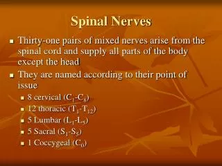
Spinal nerves • Part of the peripheral nervous system • 31 pairs attach through dorsal and ventral nerve roots • Lie in intervertebral foramina
Spinal cord segments are superior to where their corresponding spinal nerves emerge through intervetebral foramina • Spinal nerves are named according to the spinal cord segment from which they originate • 8 cervical • 12 thoracic • 5 lumbar • 5 sacral • 1 coccygeal • Cauda equina (“horse’s tail”): collection of nerve roots at inferior end of vertebral canal http://www.apparelyzed.com/spinalcord.html
Spinal nerves Dorsal roots – sensory fibers arising from cell bodies in dorsal root ganglia Ventral roots – motor fibers arising from anterior gray column of spinal cord
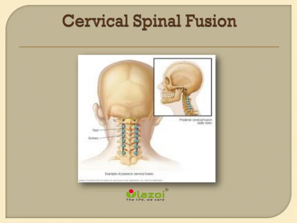
Spinal nerves • Note: cervical spinal nerves exit from above the respective vertebra • Spinal nerve root 1 from above C1 • Spinal nerve root 2 from between C1 and C2, etc. • The remaining spinal nerve pairs emerge from the spinal cord below the same-numbered vertebra • Clinically, for example when referring to disc impingement, both levels of vertebra mentioned, e.g. C6-7 disc impinging on root 7 • Symptoms usually indicate which level
Spinal nerves • Dorsal roots – sensory fibers arising from cell bodies in dorsal root ganglia • Ventral roots – motor fibers arising from anterior gray column of spinal cord (not labeled on drawing)
Dorsal and ventral roots join in an intervertebral foramen forming spinal nerve • Outside foramen, re-branch as rami (sing., ramus):Dorsal and ventral rami (somatic) Rami communicantes (visceral)
Dorsal rami serve the muscles and skin of the posterior trunk • Back, from neck to sacrum, innervated in a neatly segmented pattern: horizontal strip at same level as emergence from spinal cord • Ventral rami serve the muscles and skin of the lateral and anterior trunk • In thorax only, a simple segmented pattern as intercostal nerves • Also serve the limbs
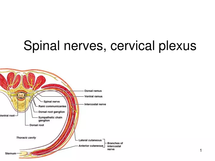
Cross section of thorax showing main roots and branches of a spinal nerve • Note dorsal and ventral roots and rami, and rami communicantes • In the thorax, each ventral ramus continues as an intercostal nerve
Nerve plexuses • Networks of successive ventral rami that exchange fibers (crisscross & redistribute) • Why would this be protective? • Mainly innervate the limbs • Thoracic ventral rami do not form nerve plexuses
Plexuses • Cervical • Brachial • Lumbar • Sacral
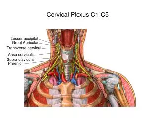
Nerve plexuses • Cervical plexus (C1-C4) innervates the muscles and skin of the neck and shoulder most important: Its phrenic nerve* (C3-C5) is the sole motor supply of diaphragm: one reason why neck injuries are so dangerous – can be lethal (respiratory arrest = stop breathing) *
Brachial plexus • Serves upper limbs and shoulder girdle • Arises primarily from C5-T1 • Main nerves (be able to label): • Musculocutaneous – to arm flexors • Median – anterior forearm muscles and lateral palm • Ulnar – anteromedial muscles of forearm and medial hand • Axillary – to deltoid and teres minor • Radial – to posterior part of limb
Sensory innervation, palm • Ulnar nerve • Median nerve • Radial nerve
Dermatomes (area of skin innervated by the cutaneous branches from a single spinal nerve is called a dermatome) Reveal sites of damage to spinal nerves or spinal cord Dermatomes (innervation of skin)