Download
1 / 45
450 likes | 808 Views
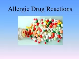
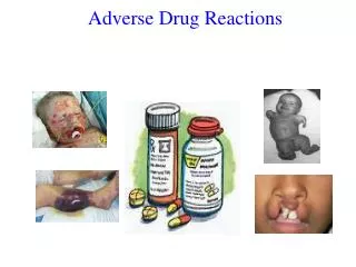
Drug Reactions. Christian Millett, MD. Overview. The skin is one of the most common targets for adverse drug reactions Exanthematous eruptions and urticaria are the two most common forms of cutaneous drug reactions The most commonly offending drugs are: penicillins, sulfonamides, and NSAIDs.

E N D
Drug Reactions Christian Millett, MD
Overview • The skin is one of the most common targets for adverse drug reactions • Exanthematous eruptions and urticaria are the two most common forms of cutaneous drug reactions • The most commonly offending drugs are: penicillins, sulfonamides, and NSAIDs
Pathogenesis • Common cutaneous drug eruptions are hypersensitivity reactions with an underlying immunologic mechanism • Type I (IgE-dependent) • Urticaria, Angioedema • Type II (Cytotoxic) • Petechiae (2o to drug-induced thrombocytopenia) • Type III (Immune complex) • Vasculitis, Serum sickness • Type IV (Delayed cell-mediated) • Exanthematous, Fixed, Lichenoid, SJS/TEN
Pathogenesis • Idiosyncratic: • AGEP (acute generalized exanthematous pustulosis) • DRESS (drug reaction with eosinophilia and systemic symptoms) • SJS/TEN • May involve a combination of immunologic interactions and genetic predisposition
Diagnosis • Most immunologically mediated drug reactions occur within 7-21 days after initiation of a new medication • Clinical characteristics • Type of primary lesion • Number and distribution of lesions • Mucous membrane involvement • Systemic signs and symptoms • Chronological factors • Drugs and dates of administration • Date of eruption • Time between drug introduction and rash • Response to removal of suspected agent
Exanthematous • Morbilliform • Polymorphous (i.e. “maculopapular”) • 7-14 days after starting a new medication • Can be earlier in cases of rechallenge • Symmetic distribution of confluent erythematous macules • Can be slightly palpable • Begins on the trunk and upper extremities • Mucous membranes are usually spared • Can be accompanied by pruritus and low-grade fever
Exanthematous • Rash disappears spontaneously after 1 to 2 weeks without sequelae • Ddx: viral exanthems • Tx: supportive • Topical steroids • Discontinue the offending agent or “treat through” • Common meds: penicillins, sulfonamides, cephalosporins, anticonvulsants
Urticaria and Angioedema • IgE-mediated immediate hypersensitivity reaction • Antigen binds to IgE on the surface of mast cells • Induces degranulation and histamine release
Urticaria • Transient erythematous, edematous papules and plaques • Associated with pruritus • Lesions can occur anywhere on the body (including palms and soles) • Individual lesions never last >24 hours • Except in the case of urticarial vasculitis
Urticaria • Most common agents: penicillins, cephalosporins, sulfonamides, tetracyclines • Tx: • Withdrawal of causative agent • H1 antihistamines
Angioedema • Edema of the deep dermis, subcutis, and submucosal tissues • Presents as swelling • Usually involves the face • Can occur anywhere from 1 day to several years after starting the drug • Most commonly due to ACE inhibitors • Can also be caused by penicillins, NSAIDs, and radiographic contrast media
Phototoxicity • Direct interaction of UV rays with the drug in the skin • Leads to generation of reaction oxygen species • Limited to sun-exposed areas • Appears as exaggerated sunburn • Most commonly due to tetracyclines, NSAIDs, and fluoroquinolones
Photoallergy • Result of cell-mediated hypersensitivity to an allergen activated or produced by the effect of light on a drug in the skin • Leads to generation of reaction oxygen species • Limited to sun-exposed areas • Lesions can resemble lichen planus • Most commonly due to thiazides and sulfonamides
Vasculitis • Typically involves small vessels • Deposition of immune complexes in postcapillary venules • Results in activation of the complement cascade • Presents as palpable purpura • Primarily on the lower extremities • Most patients only have cutaneous disease but the possibility of systemic involvement must be considered
Vasculitis • Occurs 7-21 days after drug administration • 1-3 days following rechallenge • Tx: • Withdrawal of the offending agent • Systemic steroids (if systemic involvement) • Most common agents: penicillins, NSAIDs, sulfonamides, cephalosporins
AGEP • Acute generalized exanthematous pustulosis • Presents as numerous small, sterile pustules arising within large areas of erythema • Lesions begin on face or in intertriginous zones • Accompanied by high fever • Eruption begins <2 days after drug administration • Ddx: pustular psoriasis
AGEP • Caused by: • Penicillins • Cephalosporins • Macrolides • Calcium channel blockers • Antimalarials
Fixed drug eruption • Presents as one or a few round, sharply demarcated, erythematous to dusky plaques • Common locations: face, hands/feet, genitalia • Resolves with postinflammatory hyperpigmentation • Upon rechallenge, lesions recur at the same sites
Fixed drug eruption • Caused by: sulfonamides, NSAIDs, tetracyclines
Drug-induced diseases • Sweet’s syndrome • G-CSF, all-trans-retinoic acid • Neutrophilic eccrine hidradenitis • cytarabine • Bullous pemphigoid • furosemide • Pemphigus • penicillamine, captopril
Drug-induced diseases • Psoriasis • NSAIDs, antimalarials, ACE inhibitors, beta blockers, lithium • Acne • corticosteroids, androgens, lithium, oral contraceptives, EGFR inhibitors
Drug-induced lupus • Systemic LE (SLE) • symptoms usually develop >1 year after medication is begun • associated with antihistone antibodies • caused most commonly by procainamide and hydralazine (also minocycline) • Subacute cutaneous LE (SCLE) • lesions usually occur on upper trunk and extensor arms • associated with anti-Ro and anti-La antibodies • caused most commonly by HCTZ and calcium channel blockers (also TNF-a blockers)
Drug-induced findings • Mucosal ulceration • methotrextate, doxorubicin, 5-FU • Hair loss • Telogen effluvium (anticoagulants, beta blockers, lithium) • Anagen effluvium (chemotherapeutics) • Hyperpigmentation • minocycline, antimalarials, amiodarone, bleomycin
Drug-induced findings • Injection site reactions
Drug-induced findings • Coumadin necrosis • Lesions begin 2-5 days after therapy is begun • Coincide with the early drop in protein C function • Erythematous plaques => hemorrhagic bullae and necrotic ulcers • Most common sites: breasts, thighs, buttocks • Tx: discontinue coumadin, vit K, start heparin • Heparin-induced thrombocytopenia (HIT) • Due to antibodies that bind to complexes of heparin and platelet factor 4 => platelet aggregation and consumption • Cutaneous necrosis is seen as a result of thrombosis • Tx: discontinue heparin, start argatroban
Drug-induced findings • Heparin-induced thrombocytopenia (HIT)
DRESS • Drug reaction with eosinophilia and systemic symptoms • Underlying mechanism may be alteration in metabolism of particular drugs • Most common agents are anticonvulsants and sulfonamides • Occurs 2-6 weeks after drug is begun
DRESS • Usually presents as a morbilliform eruption • Edema of the face is a frequent finding • Lymphadenopathy is often present • Prominent eosinophilia is common • Tx: • Withdrawal of the offending agent • Oral corticosteroids
SJS/TEN • Stevens-Johnson Syndrome (SJS) and Toxic Epidermal Necrolysis (TEN) are rare, life-threatening mucocutaneous diseases • Almost always drug-related • Extensive erythema and exfoliation • Due to keratinocyte cell death via apoptosis (induced by Fas-FasL interaction) => separation of skin at the dermo-epidermal junction (DEJ) • High fever and skin pain
SJS/TEN • Onset between 7-21 days after initiation of drug therapy • Can be <2 days when patients re-exposed to the drug • Immunocompromised patients at greater risk • Mortality rate: 1-5% for SJS; 25-30% for TEN
SJS/TEN • Most frequently implicated drugs: antibiotics, NSAIDs, anticonvulsants • Classified based on body surface area (BSA) • SJS: <10% of BSA • SJS-TEN overlap: 10-30% of BSA • TEN: >30% of BSA
SJS/TEN • Clinical features: • Initial symptoms can be: fever, eye pain, dysphagia • Can precede skin findings by 1-3 days • Skin lesions appear first on trunk => face, extremities • Erythematous to dusky red macules => patches • As epidermis detaches from dermis, blisters form • Positive Nikolsky sign • Skin is very painful
SJS/TEN • Erythema and erosions of the buccal, ocular, and genital mucosa
SJS/TEN • Treatment: • Immediate discontinuation of the causative drug • Supportive care • Systemic corticosteroids vs. IVIG
Take Home Points • Morbilliform eruptions and urticaria are the two most common forms of cutaneous drug reactions • The most commonly implicated drugs are: penicillins, sulfonamides, and NSAIDs • Most drug reactions occur within 7-21 days after initiation of the medication • Mucosal involvement and a positive Nikolsky sign are necessary for a diagnosis of SJS/TEN