Download
1 / 15
150 likes | 422 Views
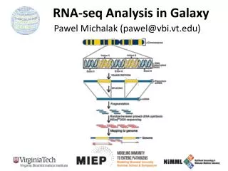
RNA-Seq. An alternative to microarray. Grow cells or isolate tissue (brain, liver, muscle) Isolate total RNA Isolate mRNA from total RNA (poly A+ select) Fragment RNA Make and amplify cDNA Sequence the ends of the cDNA Map the sequences to the human genome

E N D
RNA-Seq An alternative to microarray
Grow cells or isolate tissue (brain, liver, muscle) Isolate total RNA Isolate mRNA from total RNA (poly A+ select) Fragment RNA Make and amplify cDNA Sequence the ends of the cDNA Map the sequences to the human genome Count the number of sequence tags at each known gene (and at locations for which no gene is known) Correct for background Analyze the data Steps
PROCEDURE - Solexa –Cluster Amplification 1) Load Samples to Flow Cell 2. Attach DNA to Surface 3. Bridge Amplification 8 Lanes are loaded onto the flow cell for simultaneous analysis Single stranded DNA fragments bind randomly to the inside surface of the flow cell. Unlabeled nucleotides and enzyme are added to initiate solid-phase bridge amplification.
PROCEDURE - Solexa –Cluster Amplification 4. Fragments Become Double Stranded 5. Double Stranded Molecules are Denatured 6. Amplification is Completed The enzyme incorporates nucleotides to build double stranded bridges on the solid-phase substrate. Denaturation leaves single-stranded template anchored to the substrate Several million dense clusters of double stranded DNA are generated in each channel of the flow cell.
PROCEDURE- Solexa Sequencing & Genome Analyzer 1. Determine 1st Base 2. Image 1st Base 3. Determine 2nd Base The first sequencing cycle is initiated by adding all 4 labeled reversible terminators, primer, and DNA polymerase to the flow cell After laser excitation, an image of the emitted fluorescence from each cluster on the flow cell is captured. The 2nd sequencing cycle is initiated by adding all 4 labeled reversible terminators and enzymes.
PROCEDURE- Solexa Sequencing & Genome Analyzer 4. Image 2nd Base 5. Sequence Read Continues Over Multiple Chemistry Cycles 6. Align and Map Data After laser excitation, image data is collected like before. The identity of the 2nd base for each cluster is recorded. 35 cycles of sequencing are repeated to determine the sequence of bases in a given fragment a single base at a time. Map the sequences to the reference genome using an alignment algorithm. http://www.illumina.com/pages.ilmn?ID=204
The more mRNA copies of a specific kind, the more cDNA copies. The more cDNAs of a specific kind, the more clusters are generated on the flow cell. The more clusters are generated, the more sequence “reads” there will be for a specific mRNA. The readout is digital. Quantification Principle
MICROARRAY Grow cells or isolate tissue (brain, liver, muscle) Isolate total RNA Isolate mRNA from total RNA (poly A+ select) Make, amplify, and fluorescently label cDNA Hybridize to known complementary probes on slide array Determine the intensity of fluorescence at each spot Correct for background and normalize Analyze the data RNA-Seq Grow cells or isolate tissue (brain, liver, muscle) Isolate total RNA Isolate mRNA from total RNA (poly A+ select) Fragment RNA Make and amplify cDNA Sequence the ends of the cDNA Map the sequences to the human genome Count the number of sequence tags at each known gene (and at locations for which no gene is known) (Correct for background) and normalize Analyze the data Comparison of Steps
What can you learn with each one? What can you learn from one but not from the other? How is the primary data acquired? How are systematic biases eliminated? How do you normalize How would you look for differential expression? How would you cluster? How can you combine data from multiple experiments? Which is more sensitive? What kinds of additional software do you need? What are the similarities and differences?
Mortazavi A, Williams BA, McCue K, Schaeffer L, Wold B. Mapping and quantifying mammalian transcriptomes by RNA-Seq. Nature Methods (2008) 5: 621. http://www.illumina.com/pages.ilmn?ID=204 References