Download
1 / 54
550 likes | 589 Views
ADRENAL DISORDERS. Dr. Atallah Al-Ruhaily Conultant Endocrinolgist. Adult Adrenal Glands. Each gland weighs 4-5 g. Location: in the retroperitoneum above or medial to the upper poles of the kidneys. Surrounded by a fibrous capsule.

E N D
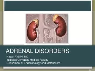
ADRENAL DISORDERS Dr. Atallah Al-Ruhaily Conultant Endocrinolgist
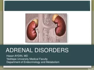
Adult Adrenal Glands • Each gland weighs 4-5 g. • Location: in the retroperitoneum above or medial to the upper poles of the kidneys. • Surrounded by a fibrous capsule. • The yellowish outer cortex comprises 90% of adrenal weight. • The inner medulla 10% of adrenal weight.
A normal right adrenal gland is shown here positioned between the liver and the kidney in the retroperitoneum. Note the amount of adipose tissue, some of which has been reflected to reveal the upper pole of the kidney and the adrenal.
Normal Adrenal Glands Each adult adrenal gland weighs from 4 to 6 grams.
Arterial Supply Adrenalcortex isrichly vascularised. Main arterial supply from branches of: • Inferior phrenic artery • Renal arteries • Aorta.
Venous Drainage • Rt. adrenal vein drains directly in the posterior aspect of I.V.C. • Lt. adrenal vein into Lt. Renal vein.
Embryology of Adrenal Cortex • Adrenal Cortex is of mesodermal origin. • Identifiable as a separate organ at the 2 month-old-fetus. • At this stage, composed of: • Fetal zone • additional zone in fetus • makes up the main bulk of weight at this time. • lost in the 1st year (usually within 3 months after birth). • Definitive zone • The origin of the adrenal cortex.
Embryology of Adrenal Cortex Fetal adrenal increases rapidly in size (Larger than the kidney at mid gestation & much larger than adult gland in relation to total body mass). Fetal adrenal produces mainly DHEA & DHEA-S (precursors of maternal-placental estrogen). Definitive zone synthesizes many steroids mainly cortisol.
Embryology of Adrenal Cortex The anatomic relationship of fetal and definitive zones is maintained until birth. Adrenocortical weight decreases gradually until the fetal zone disappears 3 months after delivery. During the first 3 years, the adult adrenal cortex develops and differentiates into 3 adult zones: Glomerulosa Fasciculata Reticularis
Embryology of Adrenal Medulla • Medullary chromaffin cells (the principal cells of adrenal medulla) are from the neural crest. • During development the medullary cells migrate and lie surrounded by the cortex.
Adrenal gland is composed of 2 distinct compartments • Adrenal cortex • produces many steroid hormones; the most important of which are: • Cortisol • Aldosterone • Adrenal androgens • Adrenal Medulla • Produces Catecholamines: • Adrenaline (Epinephrine) • Noradrenaline (Norepinephrine)
Sectioning across the adrenals reveals: • a golden yellow outer cortex; and • an inner red to grey medulla.
Microscopic Anatomy of Adrenal Gland Remember “GFR” Periadrenal Fatt Fibrous Capsule Zona Glomerulsa Zona Fasciculata Zona Reticularis Medulla
Zona Glomerulosa Zona Fasciculata Zona Reticularis Zones of Adrenal Cortex The outermost. the thickest The innermost Cells are small & lipid-poor & scattered beneath the adrenal capsule. Cells are larger, contain more lipid & termed “clear cells” “Compact” lipid-poor cells but contain lipofuscin granules. Aldosterone (Cannot produce cortisol & Androgens) Cortisol & Androgens Cortisol & Androgens Regulated by Renin-Angiotensin System & K Both structure and function are regulated by ACTH Both structure and function are regulated by ACTH Functionally considered as one unit.
Classification of Adrenal Steroids classified into 3 groups based on their predominant functions: 1. Glucocorticoids 2. Mineralocorticoids 3. Androgens
Steroid Class Androgens (C19) Glucocorticoids (C21) Mineralocorticoids (C21) Main Hormones DHEA, DHEA-S Androstenidione Cortisol Corticosterone Aldosterone Predominant action Androgenic activity • metabolism of: • carbohydrates • and proteins • metabolism of: • Na & K • Maintain the ECV Main Zone of production Z. Fasciculata Z. Reticularis (functionally as one unit) Z. Glomerulosa Major Classes of Adrenal Steroids
Disorders of Adrenal glands • Adrenal Cortex • Cushing’s syndrome • Addison’s disease • Hyperaldoteronism • Syndomes of congenital adrenal hyperplasia (CAH). • Hirsutism • Virilization Adrenal Medulla Pheochromocytoma
Adrenal Insufficiency • Adrenocortical insufficiency (hypofunction of the adrenal cortex) includes all conditions in which there is deficient production of: • adrenal glucocorticoid, and • mineralocorticoid hormones.
Types of adrenal insufficiency • These conditions are divided into 2 general groups according to the level of hypofunction: • Primary adrenal insufficiency (Addison’s diease) • due to primary hypofunction of the adrenal cortex. • Secondary adrenal insufficiency • adrenocortical failure secondary to a primary deficient secretion of ACTH from the pituitary gland.
Etiology of adrenal insufficiency Primary Adrenal Insufficiency • A. Anatomic destruction of gland (chronic & acute) • “Idiopathic” atrophy (autoimmune) • Surgical removal • Infection (Tb., fungal, viral-esp. AIDS) • Adrenal Hemorrhage • Invasion: metasteses, amyloidosis, sarcoidosis • B. Metabolic failure in hormone production • Congenital adrenal hyperplasia (CAH) • Enzyme inhibitors: • (metyrapone, ketoconazole, aminoglutethemide) • Cytotoxic agents: (mitotane) • C. ACTH-blocking Antibodies
Etiology of adrenal insufficiency Secondary Adrenal Insufficiency • A. Hypopituitarism due to hypothalamic-pituitary disease. • Suppresion of hypothalamic-pituitary axis. • Exogenous steroids (Iatrogenic) • Endogenous steroids (from tumors)
Incidence • Primary adrenal insufficiency: • relatively rare. • Occurs at any age. • affects both sexes equally. • Secondary adrenal insufficiency: • relatively common (because of common therapeutic use of steroids).
Addison’s Disease Etiology and Pathogenesis Addison’s disease results from progressive destruction of adrenal cortex. At least 90% of gland is destroyed before signs of insufficiency appear.
Addison’s Disease Etiology and Pathogenesis 50% of patients have +ve circulating adrenal Abs. Some Abs destroy the adrenal glands, others block the binding of ACTH to its receptors. Some patients have +ve Abs to thyroid, parathyroid and/or gonadal tissues. Polyglandular Autoimmune (PGA) syndromes
Endocrine Disorders Chronic lymphocytic thyroiditis Premature ovarian failure DM type 1 Primary hypothyroidism Hyperthyroidism Nonendocrine Disorders Pernicious anemia Vitiligo Alopecia Chronic active hepatitis Nontropical sprue Myasthenia gravis Associated Autoimmune Disorders
Common Symptoms in chronic primary adrenal insufficiency
Common Signs in chronic primary adrenal insufficiency
Common Laboratory findings in chronic primary adrenal insufficiency
Hyperpigmentation • Generalized hyperpigmentation of skin & mucous membrane (the classical physical finding). • Along with other features, suggests primary adrenocortical insufficiency. • One of earliest manifestations of Addison’s disease.
Hyperpigmentation • Increased at exposed areas and accentuated at pressure areas (knuckles, toes, elbows, knees) • Associated with black or dark brown freckles. • Hperpigmentation of buccal mucosa & gum is preceded by generalized hyperpigmentation of skin. • Other areas: palmar creases, nail beds, nipples, areolae, perivaginal, perianal mucosa & scars that formed after onset of ACTH excess (but not older scars).
Adrenal Imaging • Abdominal x-rays • Adrenal calcification in 50% tuberculous cases & some other invasive or hemorrhagic causes. • CT Scan more sensitive for adrenal calcification & enlargement Causes of bilateral adrenal enlargement: • Tb • Fungal infection • CMV infection • Infiltrative diseases (malignant or nonmalignant) • Adrenal hemorrhage
Acute Adrenal Crisis • A state of acute adrenal insufficiency occuring in patients with Addison’s disease who are exposed to any form of stress. • Precipitating stress factors: • Infection • Trauma • Surgery • Dehydration (Salt deprivation, vomiting, diarrhea) • Discontinuation of steroids replacement therapy
Acute Adrenal Crisis Common Clinical Features • Hypotension & shock • Fever (due to infection or hypoadrenalism per se) • Dehydration, volume depletion • Nausea, vomiting, anorexia • Abdominal pain (may mimic acute abdomen) • Weakness, apathy, depressed mentation • Hypoglycemia (more in children) • Shock and coma may rapidly lead to death in untreated patients.
Acute Adrenal Crisis Laboratory Findings Suggestive of Diagnosis • Hyponatremia & Hyperkalemia • (In a small number of acute cases). • Azotemia (usual) • Lymphocytosis • Eosinophilia • Hypoglycemia
Acute Adrenal Hemorrhage A progressively deteriorating condition resulting from bilateral adrenal hemorrhage and acute adrenal destruction in an already compromised patient with major illness.
Acute Adrenal Hemorrhage Manifestations: • Abdominal, flank or back pain & abdominal tenderness (Less frequently, abdominal distention, rigidity & rebound tenderness). • Hypotension & shock • Fever • Nausea & Vomiting • Confusion & disorientation • tachycardia
Acute Adrenal Hemorrhage With progression, the following manifestations may ensue: • severe hypotention • volume depletion • Dehydration • Hyperpyrexia • Cynosis • Hypoglycemia • Coma • death
Secondary Adrenal InsufficiencyCauses • ACTH deficiency most commonly due to exogenous glucocorticoid therapy. • Pituitary & Hypothalamus tumors the most common causes of naturally occuring pituitary ACTH hyposecretion.
Secondary Adrenal InsufficiencyPathphysiology • ACTH deficiency is the primary event. • This leads to: • decreased cortisol & androgen secretion. • But aldosterone secretion remains normal except in few cases.
Secondary Adrenal InsufficiencyPathphysiology • In early stages, • Basal ACTH & cortisol levels may be normal. • ACTH reserve is impaired. Response of ACTH & cortisol to stress is subnormal. • With further loss of basal ACTH secretion, • There is atrophy of Z. Fasciculata & Z. Reticularis. • Basal cortisol secretion is decreased • The entire pituitary adrenal axis is impaired (i.e. Decreased ACTH responsiveness to stress & decreased adrenal responsiveness to stimulation with exogenous ACTH).
Secondary Adrenal InsufficiencyClinical Features • Usually chronic nonspecific manifestations. • Acute crisis occurs in: • Undiagnosed patients • Patients who do not receive increased steroid dosage during periods of stress.
Secondary Adrenal InsufficiencyClinical Features Clinical features differ from primary in that: • Hyperpigmentation does not occur (Because of ACTH deficiency). • Manifestations of mineralocorticoid deficiency are usually absent (Because Aldosterone secretion by Z. G. is usually preserved). Therefore: • Volume depletion, dehydration & hyperkalemia usually absent. • Hypotention is usually absent except in acute presentations. • Hyponatremia may occur as a result of water retention.
Secondary Adrenal InsufficiencyClinical Features Prominent features (due to glucocorticoid deficiency) are nonspecific & include: • Weakness, lethargy & easy fatigability • anorexia, nausea & occasionally vomiting • Arthralgias & myalgias • Hypoglycemia • Acute decompensation with severe hypotention or shock unresponsive to vasopressors.
Secondary Adrenal InsufficiencyAssociated Features • The following additional features may be present: • History of glucocorticoid therapy or Cushingoid features. • Features of loss of other pituitary hormones (hypogonadism & hypothyroidism). • Features of hypersecretion of GH or PRL from pituitary adenoma. • Pressure symptoms from pituitary tumors.
Diagnosis of Adrenal Insufficiency • Basal levels of adrenocortical steroids in plasma or urine may be normal in partial adrenal insufficiency. • Tests for adrenocortical reserve are necessary to establish the diagnosis. • Rapid ACTH Stimulation Test • Plasma ACTH Levels • Metyrapone Test • Insulin-induced Hypoglycemia • CRH Stimulation
Diagnosis of Adrenal Insufficiency • Other indirect clues: • Features of hypersecretion of GH or PRL from pituitary adenoma. • Pressure symptoms from pituitary tumors.
Evaluation of Suspected Adrenal Insufficiency Rapid ACTH Stimulation Test • Abormal ACTH Stimulation Test: Adrenocortical insufficiency +ve. Which type? • Plasma ACTH level: • Elevated: Primary Adrenal Insufficiency +ve • Normal or Low: Secondary Adrenal Insufficiency +ve
Evaluation of Suspected Adrenal Insufficiency Rapid ACTH Stimulation Test • Normal ACTH Stimulation Test: • This excludes Primary Adrenal Insufficiency & Adrenal atrophy. • But does not exclude “Decreased ACTH Reserve” • Metyrapone Test • or Insulin-hypoglycemia Test • or CRH stimulation Test: • Normal: Exclude Adrenal Insufficiency • Abnormal: Secondary Adrenal Insufficiency +ve