Download
1 / 27
300 likes | 562 Views
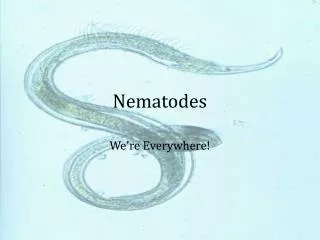
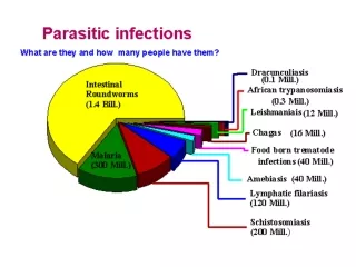
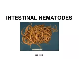
Intestinal nematodes. [4]- Enterobius vermicularis. Morphology. Pin worm - Seat worm. * Small thread-like worm, whitish in color * 2 (alae) at the anterior end * Double bulbed esophagus. Morphology cont. Male 2-5mm Curved posterior end With one spicule Female 10mm

E N D
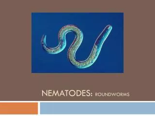
Intestinal nematodes [4]- Enterobius vermicularis
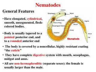
Morphology Pin worm - Seat worm * Small thread-like worm, whitish in color * 2 (alae) at the anterior end * Double bulbed esophagus
Morphology cont. Male2-5mm Curved posterior end With one spicule Female 10mm Pointed tail, 2uteri full with eggs, Vulva at the junction of Ant.¼ & post. ¾
Enterobius vermicularis Enterobiasis intestine live worm Geographical Distribution: cosmopolitan. Definitive host: man only Habitat:caecum and adjacent parts. Diagnostic stage: egg S: 50 X 20 µ ♀ 10 mm long S: plano-convex has 2 layers Plane Convex covered with outer sticky albuminous layer Eggs are found mainly on perianal skin. C: translucent C: rhabditiform larva
Mode of infection in Enterobiasis Female migrates during night ♀ ♂ Ingestion of eggs by: Autoinfection (hand to mouth) Contaminated food or drink Handling contaminated linen, clothing and articles. Air-born in dust Rhabditiform larva Retro-infection Eggs are infective in few hours
Pathogenesis and Clinical Picture • Sticky egg laid in the perianal region at night causes Pruritis ani (Perianal itching). • Pruritus ani leads to nervous irritability, hyperactivity and insomnia (أرق بالليل)
Pathogenesis and Clinical Picture Granuloma formation Fallopian tube uterus vagina urethra appendix Appendicitis Urinary infection Vulva-vaginitis Pelvic peritonitis (Involuntary micturition) Intestine Secondary enuresis diarrhoea & abdominal pain
Diagnosis Clinically: pruritus ani at night. Laboratory: -Swabbing of perianal area to detect the eggs by: NIH swab Scotch adhesive tape - Adults may be seen in stool or anal area. - Eggs are rarely found in stool NIH swab
N.I.H. swab The perianal area is swabbed in the morning before defecation with a cellophane paper tied to the tip of glass rod and inserted in a test tube . The cellophane is stretched on the slide and examined for eggs
Treatment • Albendazole. • It should be repeated after 2 weeks. • All members of the family should be simultaneously (فى نفس الوقت) treated. • Application of white mercury oxide (white precipitate ointment) around the anus at night. Relieves itching, kills female worms coming out of the anus and prevents dispersal تبعثر of eggs.
Epidemiology • Eggs become mature and infective in few hours. • Pruritus leads to autoinfection. • Prevalent in Egypt in children (5-10 years) and old age (30-35 years) . (Biphasic age group) Prevention and Control • Mass treatment. • Personal hygiene and food protection. • Infected children should use tightly fitting trousers at night.
Intestinal nematodes [5]- Ascaris lumbricoides Giant Worm
Ascaris lumbricoides Ascariasis Ascaris: worm lumbricoides: earthworm-like Geographical Distribution: cosmopolitan Green salad contaminated with embryonated Ascaris eggs 20 cm 25 cm ♀ ♂ migration Adult Ascaris worm Definitive host: Man Habitat: lumen of small intestine Ascaris eggs in stool Diagnostic stage
Diagnostic Stage Ascaris eggin stool Infective Stage Embryonated egg X Fertilized egg Decorticated egg Egg contains 2nd stage rhabditiform larva Unfertilized egg As fertilized egg but lacking mamillations S 90X40 µ 60X45 µ oval, thick smooth layer long, narrow, ill-defined mamillations S Rhabditiform Larva develops in the egg on the ground Covered by mamillations 1st moult C brownish الأرض C immature Geohelminthic infection
Development of Ascaris inside body of infected human Larva is swallowed Embryonated egg is swallowed Migration stage 2nd moult Rhabditiform larva Venous circulation Adult Ascaris in small intestine Intestinal stage 3rd & 4th moults
Pathogenesis and Clinical Picture During larval migration: Intenseشديد local inflammatory reaction around the larvae leading to pneumonitis. Eosinophilia Inflammatory cells Fever, cough, dyspnea (ضيق النفس ) and asthmatic attacks ( أزمات ربو ) and oedema of lips
Pathogenesis and Clinical Picture Adult worms in the intestine: 1- Digestive disturbances: Abdominal pain, colic, nausea, vomiting. Distension (انتفاخ) or Dyspepsia (عسر هضم) Due to production of anti-enzymes that interfere with protein digestion. Malnutrition and underdevelopment of children. Adult in the intestine 2- Changes of the bowel movements: Diarrhoea or Constipation.
مضاعفات Complications Traumatic effects: due to worm irritation by fever, drugs or anaesthesia Liver abscess Perforation, peritonitis Obstructive jaundice Appendicitis Intestinal obstruction
Intussusception a portion of the intestine enters into another portion Volvulus a portion of the intestine twists around itself
Ascaris worms enter the stomach to be vomited and may come out of external nares Ascarisworms coming out of the anus
Complications • II-Toxic effects: • caused by toxins of living or dead worms. • Oedema of face, urticaria (طفح جلدى ) , asthma (ربو ), insomnia ( أرق ), nervous irritability and convulsions Asthma Urticaria
Complications III- Larvae in ectopic sites: Larvae go to patient’s viscera Venous blood Larvae penetrated intestinal wall to blood stream Visceral larva migrans
Diagnosis Clinically: Transient cough, dyspnea (1-2 weeks). Vague غير واضحةabdominal manifestations. Laboratory: Sputum examination: streaks of blood, eosinophilia and larvae. Eosinophilia (20% - 7%) Eggs in stool. Adult in vomits, faeces. Radiologically: Plain X-ray of the lungs and Barium meal.
Plain X-ray of the lungs Normal lung Lung shows scattered mottling بقع مبعثرة Loeffler’s syndrome Barium meal
Treatment Albendazole OR Mebendazole In mixed infections: treat Ascaris infection first to prevent stimulating the worms to unwanted activity تذهب الى أماكن غير مرغوب فيها Surgical treatment of complications.
Epidemiology and Control Ascariasis is prevalent (منتشر ) at all ages, most common in preschool children. (Play and defaecate in soil) Sanitary disposal of human faeces. Nightsoil (human faeces & urine) should not be used as fertilizer for food crops. Proper washing of vegetables eaten raw. Washing of hands before meals. Mass treatment.
Case A young youth suddenly complained of abdominal pain accompanied by constipation. He was admitted to the Emergency Department of El-NOUR Hospital. He was diagnosed as a case of intestinal obstuction. a- Enumerate helminths that may cause such condition. T.solium T.saginata D.Latum ??? A.lumbricoides b- If the patient gave a history of eating Grilled beef, what is the possible parasite causing this condition? Green salad Grilled pork A.lumbricoides T.solium T.saginata c- Name the infective stage in this condition. S: 60X40µ Embryonated egg Cysticercus cellulosae C: brownish Scolex with hooks S: thick-shelled & mamillated Cysticercus bovis Bladder-like 10X5 mm Scolex with no hooks C: Larva