Download
1 / 66
770 likes | 858 Views
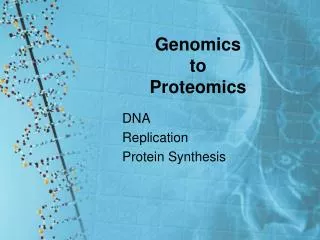
Introduction to Proteomics. Outline. What is proteomics? Why study proteins? Discuss proteomic tools and methods. What is proteomics?. Proteomics is the analysis of the protein complement to the genome. Protein. Gene. Transcript. Genomics. Proteomics.

E N D
Outline • What is proteomics? • Why study proteins? • Discuss proteomic tools and methods
Proteomics is the analysis of the protein complement to the genome Protein Gene Transcript Genomics Proteomics
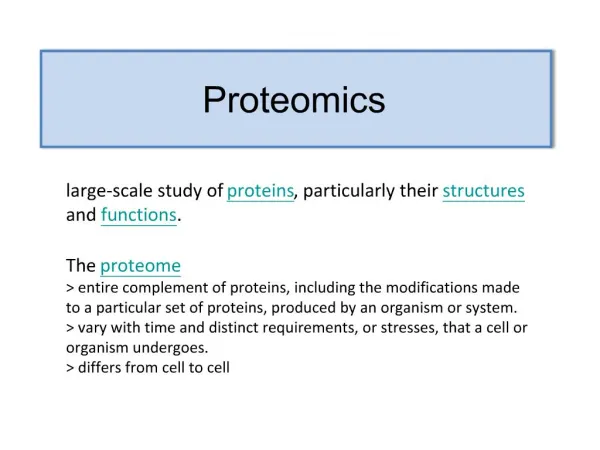
One organism will have radically different protein expression in different parts of its body, in different stages of its life cycle and in different environmental conditions.” …while the genome is a rather constant entity, the proteome differs from cell to cell and is constantly changing through its biochemical interactions with the genome and the environment. “..the large-scale study of proteins…while it is often viewed as the “next step”, proteomics is much more complicated than genomics. Wikipedia, http://en.wikipedia.org
Proteomics is multidisciplinary Protein Biochemistry Analytical Chemistry Biology Proteomics Bioinformatics Molecular Biology
Proteomics Research Basic research: To understand the molecular mechanisms underlying life. Applied research: Clinical testing for proteins associated with pathological states (e.g. cancer).
Applications of Proteomics Disease Mechanisms Medical Microbiology Signal Transduction Protein Expression Profiling Drug Discovery Glycoyslation Post-translational Modifications Proteome Mining Target ID Phosphorylation Proteolysis Differential Display Proteomics Yeast Genomics Yeast two-hybrid Protein-protein Interactions Functional Proteomics Affinity Purified Protein Complexes Co-precipitation Structural Proteomics Phage Display Mouse Knockouts Organelle Composition Subproteome Isolation Protein Complexes
For example: Hemoglobin Picks up oxygen in the lungs, travels through the blood, and delivers it to the cells. Hbα Hbβ O2 Hbα Hbβ hemoglobin
Sickle cell disease is caused by a single amino acid change. Mutated Hbβ Normal Hbβ ATG GTG CAC CTG ACT CCT GAG GAG … ATG GTG CAC CTG ACT CCT GTG GAG … … … M M V V H H L L T T P P V E E E
Summary – what is proteomics? Involves the study of proteins Proteomics is multidisciplinary Proteomics is being applied to both basic and clinical research
What are PROTEINS? Proteins are large, complex molecules that serve diverse functional and structural roles within cells.
Enzyme Defense Protease Proteins do most of the work in the cell Antibody Degrades Protein Fights Viruses O Motion Transport Actin Hemoglobin Contracts Muscles Carries O2 Regulation Insulin 2 Controls Blood Glucose Support Keratin Forms Hair and Nails
Proteins are comprised of amino acid building blocks O R1 R2 Amino acid 1 Amino acid 2 Acid O H C C O C C H C OH N O H H O H H2 N + R H C H Variable O R2 O H H2O N H H R1 C C C C H O N H H2 N H Base Dipeptide Peptide Bond
Alanine Asparagine Aspartate Cysteine Arginine Glycine Glutamate Glutamine Lysine Proline Leucine Isoleucine Methionine Serine Phenylalanine Valine Threonine Tryptophan Tyrosine Each amino acid has unique chemical properties. basic acidic Histidine non-polar hydrophobic polar hydrophilic
Proteins are chains of amino acids. O OH C N H N H H H Short chains of amino acids are called peptides. Proteins are polypeptide molecules that contain many peptide subunits.
Gene Empty tRNA Trp tRNA Ribosome Ala Trp Met Met Met Ala Met Ala tRNA tRNA Ribonucleotides A G C U Codon 1 A U G = Methionine Empty tRNA Large Subunit Codon 2 G C C = Alanine Codon 3 U G G = Tryptophan Small Subunit Codon 4 U A G = Stop 3’ Nucleus Messenger Ribonucleic Acid (mRNA) Amino Acid-transfer RNA 5’ U G G U A G A U G G C C Cytoplasm Translation is the synthesis of proteins in the cell.
Proteins have specific architecture http://www.path.cam.ac.uk/~mrc7/igs/mikeimages.html
Proteins arrive at their final structure in an ordered fashion J. E. Wampler, 1996, http://bmbiris.bmb.uga.edu/wampler/tutorial/prot0.html
Summary – why study proteins? Biological workhorses that carry out most of the functions within the cell Serve diverse functional and structural roles Composed of amino acids that are covalently linked by peptide bonds Synthesized during the translation process Must fold correctly to perform their functions
Proteomic tools to study proteins • Protein isolation • Protein separation • Protein identification
How are proteins isolated? • Mechanical Methods • grinding – break open cell • centrifugation – remove insoluble debris • Chemical Methods • detergent – breaks open cell compartments • reducing agent – breaks specific protein bonds • heat – break peptide bonds to “linearize” protein
Protein isolation procedure Grind sample in buffer Find a sample Pick it Transfer to tube Heat the sample Centrifuge to remove insoluble material “pure” protein solution Keep solution for gel analysis Recover supernatant
Protein X “pure” protein solution Isolated Protein X
Summary – protein isolation Proteins can be isolated from a variety of samples Proteomics includes the use of both mechanical and chemical methods to isolate proteins Opening cell or cellular compartments Breaking bonds and “linearizing” proteins Removal cell debris
Protein Separation SDS-PAGE
Why separate proteins? “PURE” Protein Solution Tube 1 Tube 2 Increased Complexity Decreased Complexity Decreased Protein ID Increased Protein ID
How to separate proteins? Separating intact proteins is to take advantage of their diversity in physical properties, especially isoelectric point and molecular weight
Methods of Protein Separation • Sodium Dodecyl Sulfate – Polyacrylamide Gel Electrophoresis (SDS-PAGE) • Isoelectric Focusing (IEF)
SDS-PolyAcrylamide Gel Electrophoresis (SDS-PAGE) is a widely used technique to separate proteins in solution
SDS-PAGE separates only by molecular weight • Molecular weight is mass one molecule • Dalton (Da) is a small unit of mass used to express atomic and molecular masses.
PAGE is widely used in • Proteomics • Biochemistry • Forensics • Genetics • Molecular biology
Polyacrylamide gels separate proteins and small pieces of DNA • Major components of polyacrylamide gels • Acrylamide – matrix material/ NEUROTOXIN • Bis-acrylamide - cross-linking agent/ NEUROTOXINS • TEMED - catalyst • Ammonium persulfate - free radical initiator
(matrix material) Polymerization (cross-linking agent) H H N N N H 2 a y l a i d B i s c r m e l a i d A c r y m e O O O (catalyst) N N Polyacrylamide(non-toxic) T E M E D (free radical initiator) l a A m m o n i u m p e r s u f t e S O 4
Polyacrylamide C O N H C O N H 2 2 O O N H N H Bis-acrylamide cross links C H C H 2 2 N H N H C O N H O Polyacrylamide(non-toxic) 2 O C O N H
Sodium dodecyl sulfate - SDS The anionic detergent SDS unfolds or denatures proteins • Uniform linear shape • Uniform charge/mass ratio
One-dimensional polyacrylamide gel electrophoresis (SDS-PAGE) Cathode (-) Anode (+) Standard Sample1 Sample2
During SDS-PAGE proteins separate according to their molecular weight Cathode (-) 150 kDa 100 kDa 75 kDa 50 kDa 37 kDa 25 kDa 20 kDa Bromophenol Blue dye front Anode (+) Standard Sample1 Sample2
Image of Real SDS-PAG Cathode 250 kiloDaltons 150 kDa 100 kDa 75 kDa 50 kDa 37 kDa 25 kDa 20 kDa Anode
Separation of Protein X Cathode (-) 150 kDa 100 kDa 75 kDa 50 kDa 37 kDa Protein X 25 kDa 25 kDa 20 kDa 11 kDa Bromophenol Blue dye front Anode (+) Standard Sample1 Sample2
Two-dimensional gel electrophoresis (2-DGE) 1st dimension - isoelectric focusing 2nd dimension - SDS-PAGE Most widely used protein separation technique in proteomics Capable of resolving thousands of proteins from a complex sample (i.e. blood, organs, tissue…)
1st Dimension-Isoelectric Focusing Isoelectric focusing (IEF) is separation of proteins according to native charge. isoelectric point -pH at which net charge is zero
2-DGE protein samples pH gradient 3 10 1st dimension IEF Neutral at pH 3 150 kDa 100 kDa 75 kDa 2nd dimension 50 kDa SDS-PAGE 37 kDa 25 kDa 20 kDa 11 kDa
pI kDa 3 4 5 6 7 8 9 10 100 2-DG 75 mass 50 25 Arabidopsis developing leaf
2-DGE 3 4 5 6 7 8 9 10 150 kDa 100 kDa 75 kDa 50 kDa 37 kDa 25 kDa 2nd dimension SDS-PAGE 20 kDa 11 kDa Protein X 25 kDa pI 5
1-DGE vs. 2-DGE 1-DGE (SDS-PAGE) 2-DGE Modest reproducibility Slow/Demanding Separates based on pI and size High resolution, not dependent on complexity of sample • High reproduciblity • Quick/Easy • Separates solely based on size • Modest resolution, dependent on complexity of sample
Summary – protein separation Protein separation takes advantage physical properties such as isoelectric point and molecular weight SDS-PAGE is a widely used technique to separate proteins 1-DGE is a quick and easy method to separate protein by size only 2-DGE combines isoeletric focusing (IEF) and SDS-PAGE to separate proteins by pI and size