Download
1 / 13
130 likes | 148 Views
Explore the structure of a long bone (humerus) through detailed diagrams and descriptions in this quiz designed for Anatomy & Physiology students. Study the different components such as spongy bone, compact bone, medullary cavity, and more. Gain a deeper understanding of the microscopic structure of compact bone, including osteons and lamellae. Visualize the human skeleton, highlighting the major bones and their positioning. Perfect for students of H. Jefferson Township High School. Instructor: Phyllis Smith. Information on Quiz - Ch. 5 – Pages 134-144.

E N D
Diagrams for Bones: An Overview Quiz Anatomy & Physiology H Jefferson Township High School Instructor: Phyllis Smith Information on Quiz - Ch. 5 – Pages 134 - 144
The structure of a long bone (humerus). (a) Anterior view with longitudinal section cut away. Figure 1
Figure 5.2a The structure of a long bone (humerus). (a) Anterior view with longitudinal section cut away at the proximal end. Spongy bone Proximal epiphysis Articular cartilage Epiphyseal line Periosteum Compact bone Medullary cavity Diaphysis Distal epiphysis (a)
The structure of a long bone (humerus). Pie shaped 3D view. Figure 2
Figure 5.2b The structure of a long bone (humerus). (b) Pie-shaped, three-dimensional view of spongy bone and compact bone of the epiphysis. Spongy bone Compact bone Articular cartilage (b)
The structure of a long bone (humerus). Cross section of the shaft (diaphysis). Figure 3
Figure 5.2c The structure of a long bone (humerus). (c) Cross section of the shaft (diaphysis). Endosteum Yellow bone marrow Compact bone Periosteum Perforating fibers Nutrient arteries (c)
Figure 5.3a Microscopic structure of compact bone. (a). Osteon (Haversian system) Lamellae Blood vessel continues into medullary cavity containing marrow Spongy bone Perforating fibers Compact bone Periosteal blood vessel Central (Haversian) canal Periosteum Perforating (Volkmann’s) canal (a) Blood vessel
Microscopic structure of compact bone. Highly magnified Figure 5
Figure 5.3b Microscopic structure of compact bone. (b). Lamella Osteocyte Canaliculus Lacuna Central (Haversian) canal (b)
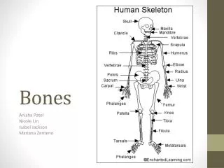
The human skeleton. (a) Anterior view. Figure 6 (a) Anterior view
Figure 5.6a The human skeleton. (a) Anterior view. Cranium Skull Facial bones Clavicle Thoracic cage (ribs and sternum) Scapula Sternum Rib Humerus Vertebra Vertebral column Radius Ulna Sacrum Carpals Phalanges Metacarpals Femur Patella Tibia Fibula Tarsals Metatarsals Phalanges (a) Anterior view