Download
1 / 48
480 likes | 582 Views
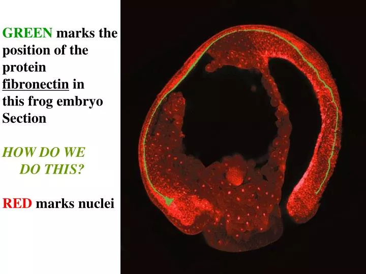
GREEN marks the position of the protein fibronectin in this frog embryo Section HOW DO WE DO THIS? RED marks nuclei. Making Polyclonals serum + or - purif. MCB 6.2 ( ‘ 0601 ’ ) Monoclonals. PAGE PolyAcrylimide Gel Elecrophoresis MCB 3

E N D
GREEN marks the position of the protein fibronectin in this frog embryo Section HOW DO WE DO THIS? RED marks nuclei
Making Polyclonals serum + or - purif.
MCB 6.2(‘0601’) Monoclonals
PAGE PolyAcrylimide Gel Elecrophoresis MCB 3 Western or Immunoblot MCB 3.5 “3EIMMBLOT”
Immunocytochemistry Most common enzyme conjugates: Alkaline phosphatase Horseradish Peroxidase
Fluorescent microscopy FITC secondary
Anti-fibronectin Then FITC Fluorescence, rather than a converted substrate, as secondary to mark protein’s presence RED, PI, nuclear counterstain
Confocal – What it offers Fluorescence microscopy Regular Confocal
Hunchback Kruppel
Immunogold SO: Immunogold Immunofluorescence Immunocytochemistry
Tracking specific macromolecules For protein: antibody-antigen For nucleic acid: n.a. complementarity
MCB 7.2 PCR Start here week2/3
In situ hybridization using radioactive probe -expose photographic emulsion
Northern Northern or SLOT-BLOT
Northern - or dot/slot blot Developmental Northern or SLOT-BLOT Microarray Tiling Microarray RNA seq. Single cell RNA seq. RIBOSOME PROFILING- on way to proteome
Figure 4.16(1) Microarray Analysis of Those Genes Whose Expression in the Early Xenopus Embryo Is Caused by the Activin-Like Protein Nodal-Related 1 (Xnr1)
Figure 4.16(2) Microarray Analysis of Those Genes Whose Expression in the Early Xenopus Embryo Is Caused by the Activin-Like Protein Nodal-Related 1 (Xnr1)
Northern - or dot/slot blot Developmental Northern or SLOT-BLOT Microarray Tiling Microarray RNA seq. Single cell RNA seq. RIBOSOME PROFILING- on way to proteome
MCB 5.1 Reporter Constructs
Myf-5 Driven Beta-gal X-gal
GFP spindles http://www.duke.edu/web/microlabs/endow/moviepage.html
Antibody, FITC to P granules Hoescht-Dye