Download
1 / 6
150 likes | 665 Views
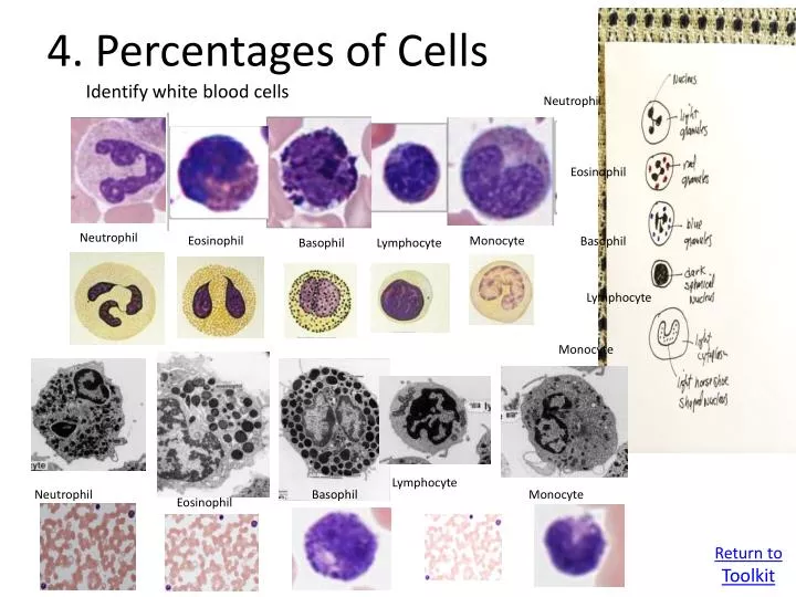
4 . Percentages of Cells. Identify white blood cells . Neutrophil. Eosinophil. Neutrophil. Eosinophil. Monocyte. Basophil. Basophil. Lymphocyte. Lymphocyte. Monocyte. Lymphocyte. Neutrophil. Basophil. Monocyte. Eosinophil. Return to Toolkit. Eosinophil. Neutrophil. Basophil.

E N D
4. Percentages of Cells Identify white blood cells Neutrophil Eosinophil Neutrophil Eosinophil Monocyte Basophil Basophil Lymphocyte Lymphocyte Monocyte Lymphocyte Neutrophil Basophil Monocyte Eosinophil Return to Toolkit
Eosinophil Neutrophil Basophil Lymphocyte Monocyte
Size of human blood cells Cell/plateletsize 1. Erythrocytes 6.5-8 µm 2. Leukocytes (WBC) % of WBC a) Neutrophil 12-15 µm 60-70% b) Eosinophil 12-15 µm 2-4% c) Basophil 12-15 µm 0-1% d) Lymphocyte 6-18 µm 25% e) Monocyte 12-20 µm 5% 3. Platelets 2-4 µm
Human blood cellsand functions Cell type Main functions Erythrocyte CO2 and O2 transport Neutrophil phagocytosis of bacteria Eosinophil parasitic infections, inflammatory processes Basophil release of histamine and other inflammation mediators Monocyte Mononuclear-phagocyte system become macrophages
Human blood cellsand functions con’t Cell typemain functions B lymphocytes generation of antibody-producing plasma cells T lymphocytes killing of virus-infected cells Natural killer killing of some tumor and (cytotoxic T cell) virus-infected cells Platelets clotting of blood
1. Operating Microscope Images Online Microscope Image Links: Healthy Blood Smear Image Unhealthy Blood Smear Image Human Testes Image To move around the image, click and drag with the mouse to move to different parts of the image. To move quickly to a completely new area, click at one corner and drag to the opposite corner. Note: These images are taken from dead but preserved cells and tissues which were stained to allow observation and digitizing by light or electron microscopy. Return to Toolkit