Download
1 / 45
650 likes | 2.25k Views
Photoreception - Vision. Vision. Accessory structures of the eye. Eyelids ( palpebrae ) separated by the palpebral fissue Eyelashes Tarsal glands Lacrimal apparatus. The eye can only perceive a small portion of the spectrum of electromagnetic waves. Vision. In order to see an object:

E N D
Vision Accessory structures of the eye • Eyelids (palpebrae) separated by the palpebralfissue • Eyelashes • Tarsal glands • Lacrimal apparatus
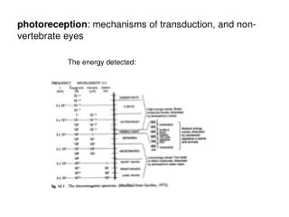
The eye can only perceive a small portion of the spectrum of electromagnetic waves
Vision • In order to see an object: - 1- the pattern of the object must fall on the vision receptors (rods and cones in the retina) accommodation - 2- the amount of light entering the eye must be regulated (too much light will “bleach out” the signals) - 3- the energy from the waves of photons must be transduced into electrical signals - 4- The brain must receive and interpret the signals
External Structures of the Eye • Conjunctiva covers most of eye • Cornea is transparent anterior portion
Lacrimal Apparatus • Secretions from the lacrimal gland contain lysozyme • Tears form in the lacrimal glands, wash across the eye and collect in the lacrimal lake • Pass through the lacrimalpunctae, lacrimalcanaliculi, lacrimal sac and nasolacrimal duct
The eye • Three layers • Outer fibrous tunic • Sclera, cornea, limbus • Middle vascular tunic • Iris, ciliary body, choroid • Inner nervous tunic • Retina
Internal Structures of the Eye • Ciliary body • Ciliary muscles and ciliary processes, which attach to suspensory ligaments of lens • Retina • Outer pigmented portion • Inner neural part • Rods and cones
The iris controls the amount of light entering the eye cavities The contraction of radial or circular smooth muscles located within the iris permit changes in the pupil diameter Regulation of the Amount of Light Entering the Eye
Retina • Retina contains rods and cones • Cones densely packed at fovea (center of the macula lutea) • Retinal pathway • Photoreceptors to bipolar cells to ganglion cells, to the brain via the optic nerve • Axons of ganglion cells converge at blind spot (optic disc) • Horizontal cells and amacrine cells modify the signal passed along the retinal neurons
Three cell layers: -- outer layer: photoreceptors- rods and cones -- middle layer: bipolar neurons -- inner layer: ganglion cells Retinal structure
Eye Anatomy • Ciliary body and lens divide the anterior cavity of the eye into posterior (vitreous) cavity and anterior cavity • Anterior cavity further divided • anterior chamber in front of eye • posterior chamber between the iris and the lens
Fluids in the eye • Aqueous humor circulates within the eye • diffuses through the walls of anterior chamber • passes through canal of Schlemm • re-enters circulation • Vitreous humor fills the posterior cavity. • Not recycled – permanent fluid
Glaucoma Cataract Eye Abnormalities
Lens • Posterior to the cornea and forms anterior boundary of posterior cavity • Posterior cavity contains vitreous humor • Lens helps focus • Light is refracted as it passes through lens • Accommodation is the process by which the lens adjusts to focus images • Normal visual acuity is 20/20
Accommodation • It is the process of adjusting the shape of the lens so that the external image fall exactly on the retina
Accommodation Abnormalities • Myopia • Hyperopia • Astigmatism: the cornea is irregular irregular pattern of vision • Presbyopia: stiffening of the lens occurring with aging increased difficulty with near vision
Visual Physiology • Rods – respond to almost any photon • Cones – specific ranges of specificity
Photoreceptor Structure • Outer segment with membranous discs • Narrow stalk connecting outer segment to inner segment • Light absorption occurs in the visual pigments • Derivatives of rhodopsin
Photons hit the pigment of a photoreceptor enzymes are activated in the cell which modify its state of polarization the signals are sent to visual area of the occipital lobe of the brain through the optic nerve Phototransduction - General
Color sensitivity • Integration of information from red, blue and green cones • Colorblindness is the inability to detect certain colors
Retinal Adaptation • Dark adapted – most visual pigments are fully receptive to stimulation • Light adapted – pupil constricts and pigments bleached.
The Visual Pathway • Large M-cells monitor rods • Smaller more numerous P cells monitor cones
Seeing in Stereo • Vision from the field of view transfers from one side to the other while in transit • Depth perception is obtained by comparing relative positions of objects from the two eyes
Neural processing • The bipolar neurons and ganglion cells process the signal • In the fovea where the acuity is the highest: 1 cone 1 bipolar cell 1 ganglion cell • At the periphery: many rods 1 bipolar cell … acuity is much decreased • Other cells in the retina participate in signal processing
Visual Circadian Rhythm • Input to suprachiasmic nucleus affects the function of the brainstem • Circadian rhythm ties to day-night cycle, and affects metabolic rates