Download
1 / 45
450 likes | 678 Views
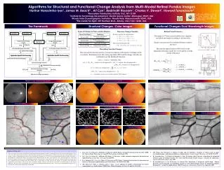
Structural Images. 杜政昊 Cheng- Hao Tu , PhD. Outlines. Computational Anatomy Voxel-based Morphometry (VBM) Diffusion Tensor Imaging (DTI). Aims of computational neuroanatomy. Many interesting and clinically important questions might relate to the shape or local size of regions of the brain

E N D
Structural Images 杜政昊Cheng-HaoTu, PhD
Outlines • Computational Anatomy • Voxel-based Morphometry (VBM) • Diffusion Tensor Imaging (DTI)
Aims of computational neuroanatomy • Many interesting and clinically important questions might relate to the shape or local size of regions of the brain • For example, whether (and where) local patterns of brain morphometry help to: • Distinguish schizophrenics from healthy controls • Understand plasticity, e.g. when learning new skills • Explain the changes seen in development and aging • Differentiate degenerative disease from healthy aging
Computational Neuroanatomy Baseline Image Standard clinical MRI 1.5T T1 SPGR 1x1x1.5mm voxels Repeat image 12 month follow-uprigidly registered Subtraction image
Why we use VBM • The output of VBM can be either information concerning regional volume or tissue concentration (density) • VBM has been designed to be sensitive to differences in local compositions of various brain tissue types, such as gray matter • The output of VBM can be displayed as a statistical parametric map
Short history of VBM A Voxel-Based Method for the Statistical Analysis of Gray and White Matter Density… Wright, McGuire, Poline, Travere, Murrary, Frith, Frackowiak and Friston. NeuroImage 2(4), 1995 (!) Rigid reorientation (by eye), semi-automatic scalp editing and segmentation, 8mm smoothing, SPM statistics, global covars. Voxel-Based Morphometry – The Methods. Ashburner and Friston. NeuroImage 11(6 pt.1), 2000 Non-linear spatial normalisation, automatic segmentation Thorough consideration of assumptions and confounds
Short history of VBM A Voxel-Based Morphometric Study of Ageing…Good, Johnsrude, Ashburner, Henson and Friston. NeuroImage 14(1), 2001 Optimised GM-normalisation (“a half-baked procedure”), modulation of segments with Jacobiandeterminants Unified Segmentation. Ashburner and Friston. NeuroImage 26(3), 2005 Principled generative model for segmentation usingdeformable priors A Fast Diffeomorphic Image Registration Algorithm. Ashburner. Neuroimage 38(1), 2007 Large deformation normalisation to average shape templates
Preprocessing – General steps • Segmentation: separate tissue types • Spatial normalization: corrects for global differences in position and transform to stereotactic space • (Modulation): keep volume information • Smooth: modifying the data to fit a certain distribution for statistical analysis.
Segmentation- basic approach • Intensities are modelled by a Gaussian Mixture Model • Parameterised by means, variances and mixing proportions
Segmentation – modified approach • Multiple components per tissue class allow non-Gaussian distributions to be modelled • accounting for partial volume effects
Tissue Probability Maps • Tissue probability maps (TPMs) can be used to provide a spatially varying prior distribution, which is tuned by the mixing proportions GMWMCSF Other
Image Registration • Registration - i.e. Optimise the parameters that describe a spatial transformation between the source and reference (template) images • Transformation - i.e. Re-sample according to the determined transformation parameters
Optimisation • Optimisation involves finding some “best” parameters according to an “objective function”, which is either minimised or maximised • The “objective function” is often related to a probability based on some model Most probable solution (global optimum) Objective function Local optimum Local optimum Value of parameter
Spatial normalization • Estimate the best 12-parameter affine transformation. • Correction for non-linear, global differences. • A mask weights the normalization to brain instead of non-brain. • Reslice the images. Original Warped Template
Modulation Native 1 1 • Multiplication of the warped (normalised) tissue intensities so that their regional or global volume is preserved • Can detect differences in completely registered areas Unmodulated 1 1 1 1 Modulated 2/3 1/3 1/3 2/3 Original Warped Modulated
Smoothing • The analysis will be most sensitive to effects that match the shape and size of the kernel • The data will be more Gaussian and closer to a continuous random field for larger kernels • Results will be rough and noise-like if too little smoothing is used • Too much will lead to distributed, indistinct blobs
Smoothing • Between 7 and 14mm is probably reasonable • (DARTEL’s greater precision allows less smoothing) • The results below show two fairly extreme choices, 5mm on the left, and 16mm, right
Grey matter Grey matter Grey matter Grey matter White matter White matter White matter White matter Simultaneous registration of GM to GM and WM to WM Subject 1 Subject 3 Grey matter White matter Template Subject 2 Subject 4
Mis-register Mis-classify Folding Thinning Mis-register Thickening Mis-classify Interpreting findings
“Globals” for VBM • Shape is really a multivariate concept • Dependencies among volumes in different regions • SPM is mass univariate • Combining voxel-wise information with “global” integrated tissue volume provides a compromise • Using either ANCOVA or proportional scaling (ii) is globally thicker, but locally thinner than (i) – either of these effects may be of interest to us.
Total Intracranial Volume (TIV/ICV) • “Global” integrated tissue volume may be correlated with interesting regional effects • Correcting for globals in this case may overly reduce sensitivity to local differences • Total intracranial volume integrates GM, WM and CSF, or attempts to measure the skull-volume directly • Not sensitive to global reduction of GM+WM (cancelled out by CSF expansion – skull is fixed!) • Correcting for TIV in VBM statistics may give more powerful and/or more interpretable results
VBM’s statistical validity • Residuals are not normally distributed • Little impact on uncorrected statistics for experiments comparing reasonably sized groups • Probably invalid for experiments that compare single subjects or tiny patient groups with a larger control group • Mitigate with large amounts of smoothing • Or use nonparametric tests that make fewer assumptions, e.g. permutation testing with SnPM
VBM’s statistical validity • Correction for multiple comparisons • RFT correction based on peak heights is fine • Correction using cluster extents is problematic • SPM usually assumes that the smoothness of the residuals is spatially stationary • VBM residuals have spatially varying smoothness • Bigger blobs expected in smoother regions • Cluster-based correction accounting for nonstationary smoothness is under development • Or use SnPM…
VBM’s statistical validity • False discovery rate • Less conservative than FWE • Popular in morphometric work • (almost universal for cortical thickness in FreeSurfer) • Recently questioned… • Topological FDR (for clusters and peaks) • See SPM8 release notes and Justin’s papers • http://dx.doi.org/10.1016/j.neuroimage.2008.05.021 • http://dx.doi.org/10.1016/j.neuroimage.2009.10.090
Longitudinal VBM • The simplest method for longitudinal VBM is to use cross-sectional preprocessing, but longitudinal statistical analyses • Standard preprocessing not optimal, but unbiased • Non-longitudinal statistics would be severely biased • (Estimates of standard errors would be too small) • Simplest longitudinal statistical analysis: two-stage summary statistic approach (common in fMRI) • Within subject longitudinal differences or beta estimates from linear regressions against time
Diffusion Tensor Imaging (DTI) • Non invasive way of understanding brain structural connectivity • Macroscopic axonal organization • Contrast based on the directional rate of diffusion of water molecules
Imaging • MR signal is attenuated as a result of diffusion • Brownian motion in the presence of gradients • Gradients are applied in different directions and attenuation is measured • DTI reconstruction using linear system solution
Effect of Varying Gradient Direction DWI z DWI x DWI y
What is b? • b-value gives the degree of diffusion weighting and is related to the strength and duration of the pulse gradient as well as the interval between the gradients • b changes by lengthening the separation of the 2 gradient pulses more time for water molecules to move around more signal loss (imperfect rephasing) • G= gradient amplitude • δ = duration • = trailing to leading edge separation
Diffusion Tensor • Mobility in a given direction is described by ADC • The tissue diffusivity is described by the tensor D • The diffusion equation • Diffusion is represented by a 33 tensor matrix* *P. Basser and D. Jone, NMR in Biomedicine, 2002.
`Diffusion MRI` Johansen-Berg and Behrens
l1+l2+l3 MD = <l> = 3 Indices of Diffusion Simplest method is the MEAN DIFFUSIVITY (MD): • This is equivalent to the orientationally averaged mean diffusivity
Indices of Anisotropic Diffusion • Fractional anisotropy (FA): • The calculated FA value ranges from 0 – 1 : FA= 0 → Diffusion is spherical (i.e. isotropic) FA= 1 → Diffusion is tubular (i.e. anisotropic)
Color FA Map • Single Tensor estimation • Estimation of direction is severely affected in the presence of noise* *X. Ma, Y. Kadah, S. LaConte, and X. Hu, Magnetic Resonance in Medicine, 2004.
Source of noise in diffusion MRI • Statistical Noise • Magnetic Filed inhomogeneity • Eddy currents • Thermal Noise • background signals caused by processing tissue magnetization. • Systematic Noise • from a number of patient motion, such as respiration, vascular, and CSF pulsations; • receiver-coil or gradient-coil motion; aliasing; and data truncation (Gibbs) artifacts
DTI-Based Connectivity Mapping • Nerve fibers represent cylindrical-shaped physical spaces with membrane acting like a barrier • DT shows diffusion preference along axon • Measuring the diffusing anisotropy, we can estimate the dominant direction of the nerve bundle passing through each voxel Johansen-Berg et al. Ann Rev. Neurosci 32:75-94 (2009)
Tractography – Techniques Degree of anisotropy Streamline tractography Probabilistic tractography Nucifora et al. Radiology 245:2 (2007)
Streamline (deterministic) tractography • Connects neighbouring voxels from user defined voxels (SEED REGIONS) e.g. M1 for the CST • User can define regions to restrict the output of a tract e.g. internal capsule for the CST • Tracts are traced until termination criteria are met (e.g. anisotropy drops below a certain level or there is an abrupt angulation)
Probabilistic tractography • Value of each voxel in the map = the probability the voxel is included in the diffusion path between the ROIs • Run streamlines for each voxel in the seed ROI • Provides quantitative probability of connection at each voxel • Allows tracking into regions where there is low anisotropy e.g. crossing or kissing fibres
Crossing/Kissing fibres Crossing fibres Kissing fibres Low FA within the voxels of intersection
Crossing/Kissing fibres Assaf et al. J Mol Neurosci 34(1) 51-61 (2008)
DTI - Tracts Corticospinal Tracts - Streamline Corticospinal Tracts -Probabilistic Nucifora et al. Radiology 245:2 (2007)