Download
1 / 54
570 likes | 960 Views
OSTEOLOGY. YENI DHAMAYANTI. GENERAL OBJECTIVES :. The students understands about structure and position of the bones which formed the limbs and they articulations. Specific Objectives :. The Students are know about : Structure, location and content of bones of the thoracic appendage

E N D
OSTEOLOGY YENI DHAMAYANTI
GENERAL OBJECTIVES : The students understands about structure and position of the bones which formed the limbs and they articulations
Specific Objectives : The Students are know about : • Structure, location and content of bones of the thoracic appendage • Structure, location and content of bones of the pelvic limb • Articulation at the thoracic and pelvic limb
The Skeleton may be divided primarily into three parts : • THE AXIAL SKELETON; comprises the vertebral column, ribs, sternum and skull. • THE APPENDICULAR SKELETON; incudes the bones of the limbs. • THE VISCERAL or SPLECHNIC SKELETON; consits of certain bones developed in the substance of some of the viscera or soft organs, e.g. os penis of the dog and os cordis of the ox
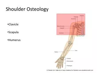
OSSA APPENDICULARIS • BONES OF THE THORACIC LIMB = OSSA MEMBRI THORACICI (EXTREMITAS CRANIALIS) • BONES OF THE PELVIC LIMB = OSSA MEMBRI PELVINA (EXTREMITAS CAUDALIS)
The thoracic limb of animals are composed of four chief segments : THE THORACIC GIRDLE REGIO CINGULUM MEMBRI THORACICI OS. CLAVICULA OS. SCAPULA OS. CORACOIDEUS
The thoracic girdle attaches the forelimb to the body and is incomplete in domestic mamals. A complete pectoral girdle consists a scapula, coracoids and clavicles Climbing and burrowing mamals usually posess a scapula and clavicle, coursing and grazing mamals usually posess a scapula only. All three pairs of bones of the thoracic girdle are seen in birds and reptiles.
THE ARM REGIO BRACHII OS HUMERUS
THE FOREARM REGIO ANTEBRACHII OS RADIUS OS ULNA
THE MANUS REGIO MANUS OSSA CARPI OSSA PHALANX OS METACARPI OSSA SESSAMOIDEA
OS SCAPULA (facies lateralis) SPINA SCAPULA FOSSA SUPRASPINATA TUBER SPINA FOSSA INFRASPINATA COLLUM SCAPULA THE HORSE
CARTILAGO SCAPULA ANGULUS CRANIALIS ANGULUS CAUDALIS ANGULUS GLENOIDALES
DISTAL OS SCAPULA TUBERCULUM SUPRAGLENOIDEUS PROCESSUS CORACOIDEUS INCISSURA GLENOIDALES CAVITAS GLENOIDALES
OS HUMERUS • EXTREMITAS PROXIMALIS • CORPUS • EXTREMITAS DISTALIS
EXTREMITAS PROXIMAL HUMERUS (DORSAL VIEW) TUBERCULUM MINOR TUBERCULUM MAJOR TUBERCULUM INTERMEDIUS CAPUT HUMERI
The comparative of the proximal extremity of the horse and the cattle
Proximal Extremity Ruminant • Lateral tuberosity is very large, and rises abour 3 cm proximal to the level of the head, forming the point of the shoulder. • Its cranial part curves medially over the intertuberal groove, and distal to it laterally there is a prominent circular rough area for the insertion of the tendon of the infraspinatus muscle.
CORPUS HUMERI (horse) TUBEROSITAS DELTOIDEUS SULCUS BRACHIALIS
CORPUS HUMERI KUDA TUBEROSITAS TERES MAJOR
EXTREMITAS CAUDALIS OS HUMERUS FOSSA OLECRANON TROCHLEA CAPITULUM
EXTREMITAS CAUDALIS OS HUMERUS EPICONDYLUS MEDIALIS CRISTA EPICONDYLOIDEA LATERALIS EPICONDYLUS LATERALIS P.T.O. EXTENSOR
EXTREMITAS CAUDALIS OS HUMERUS KUDA FOSSA OLECRANON FOSSA RADIALIS
OS RADIUS & OS ULNA OS ULNA OS RADIUS
OS RADIUS FOVEA CAPITULARIS CORPUS OS RADIUS TROCHLEA RADIALIS
OS ULNA KUDA OLECRANON PROC. ANCONEUS SPATIUM INTEROSSEUM ANTEBRACHII INCISURA SEMILUNARIS
OS ULNA SAPI SPATIUM INTEROSSEUS ANTEBRACHII
OS ULNA SAPI PROCESSUS STYLOIDEUS LATERALIS
OSSA CARPALES • The capus consists of a group of six to eight bones, depending on the species of animal. • The bones arranged in two rows, proximal and distal.
The accessory carpal bone is situated to palmar to the ulnar carpal bone and the lateral part of the trochlea of the radius. It is discoid and the medial surface is form the lateral wall of the carpal groove (canalis carpalis).
OSSA METACARPAL FACIES ARTICULARIS IV III CORPUS MC II TROCHLEA MC
OSSA METACARPAL V SLV SLD III IV INCISSURA INTERTROCHLEARIS
OSSA DIGITORUM MANUS COMPEDALE CORONALE UNGULARE
OSSA DIGITORUM MANUS OSSA SESAMOIDEA PROXIMALIS OSSA SESAMOIDEA DISTALIS
THANK YOU FOR YOUR ATTENTION
TUBEROSITAS TERES MAJOR Middle of the medial surface of the shaft is a small roughened, which the conjoined tendon of the latissimus dorsi and teres major muscles is atteched.
SULCUS BRACHIALIS • = SULCUS MUSCULOSPIRALIS • The lateral surface is smooth and is spirally curved, which contains the brachialis muscles. • RUMINANSIA, KARNIVORA and PIG is shallow.
TUBEROSITAS DELTOIDEUS Cranial surface and lateral surface are separated by a distinct border, the crest of the humerus, which bears proximal to its middle the deltoid tuberosity, to which the deltoideus muscle inserts.
CAPUT HUMERI The head presents an almost circular convex articular surface, which is about twice as extensive as the glenoid cavity of the scapula, with which it articulates and IT IS POSITION AT THE CAUDALof the proximal extremity.
TUBERCULUM HUMERI • The greater tubercle = tuberculum major (lateral tuberosity) is placed craniolaterally • The lesser tubercle = tuberculum minor (medial tuberosity) is placed craniomedially • The intertuberal or bicipital groove is bounded by the cranial parts of both tubercles, and is subdivided by and intermediate tubercle or ridge.
TUBERCULUM INTERMEDIUS • The third tubercle at median • Look clearly at horse, but we can see clear at ruminantia, carnivora and pig • This tubercle divided from major et minor by the groove, SULCUS BICIPITIS or SULCUS INTERTUBELARIS • The groove, in fresh, lodges the tendon of origin of the biceps brachii muscle.