Download
1 / 25
250 likes | 264 Views
This chapter explores the autonomic nervous system (ANS), including its regulation of smooth muscle, cardiac muscle, and certain glands. It delves into the structures involved in the ANS, such as the general visceral afferent and efferent neurons, and the integration center within the brain. Additionally, it discusses the differences between the autonomic and somatic nervous systems and provides an overview of the basic anatomy of the ANS, including the preganglionic and postganglionic neurons. The chapter also explores the two major divisions of the ANS, the parasympathetic and sympathetic divisions, as well as their sources of dual innervation and the locations of autonomic ganglia. Furthermore, it covers the autonomic plexuses and the structures and pathways of the sympathetic and parasympathetic nervous systems. The chapter concludes with a discussion on the neurotransmitters classified as cholinergic or adrenergic neurons within the ANS.

E N D
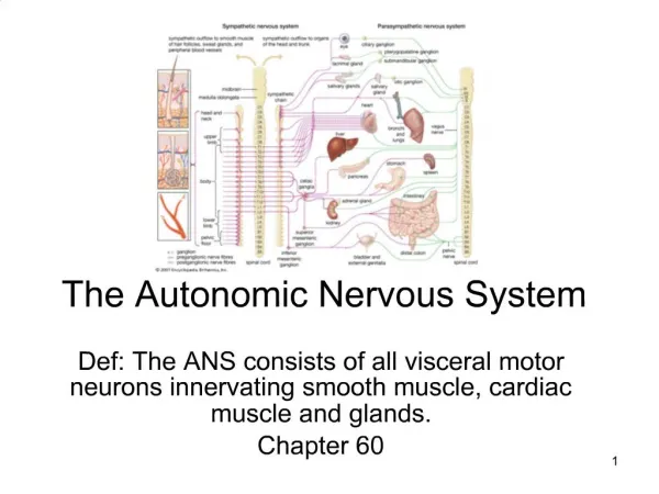
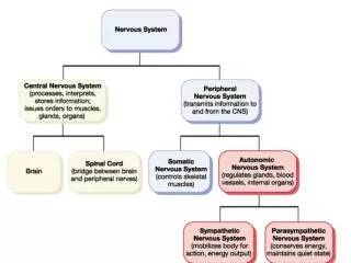
Chapter 17 The Autonomic Nervous System • Regulate activity of smooth muscle, cardiac muscle & certain glands • Structures involved • general visceral afferent neurons • general visceral efferent neurons • integration center within the brain • Receives input from limbic system and other regions of the cerebrum
Autonomic versus Somatic NS • Somatic nervous system • consciously perceived sensations • excitation of skeletal muscle • one neuron connects CNS to organ • Autonomic nervous system • unconsciously perceived visceral sensations • involuntary inhibition or excitation of smooth muscle, cardiac muscle or glandular secretion • two neurons needed to connect CNS to organ • preganglionic and postganglionic neurons
Autonomic versus Somatic NS • Notice that the ANS pathway is a 2 neuron pathway while the Somatic NS only contains one neuron.
Basic Anatomy of ANS • Preganglionic neuron • cell body in brain or spinal cord • axon is myelinated type B fiber that extends to autonomic ganglion • Postganglionic neuron • cell body lies outside the CNS in an autonomic ganglion • axon is unmyelinated type C fiber that terminates in a visceral effector
Divisions of the ANS • 2 major divisions • parasympathetic • sympathetic • Dual innervation • one speeds up organ • one slows down organ • Sympathetic NS increases heart rate • Parasympathetic NS decreases heart rate
Sources of Dual Innervation • Sympathetic (thoracolumbar) division • preganglionic cell bodies in thoracic and first 2 lumbar segments of spinal cord • Parasympathetic (craniosacral) division • preganglionic cell bodies in nuclei of 4 cranial nerves and the sacral spinal cord
Locations of Autonomic Ganglia • Sympathetic Ganglia • trunk (chain) ganglia near vertebral bodies • prevertebral ganglia near large blood vessel in gut • celiac • superior mesenteric • inferior mesenteric • Parasympathetic Ganglia • terminal ganglia in wall of organ
Autonomic Plexuses • Cardiac plexus • Pulmonary plexus • Celiac (solar) plexus • Superior mesenteric • Inferior mesenteric • Hypogastric
Structures of Sympathetic NS • Preganglionic cell bodies at T1 to L2 • Rami communicantes • white ramus = myelinated = preganglionic fibers • gray ramus = unmyelinated = postganglionic fibers • Postganglionic cell bodies • sympathetic chain ganglia along the spinal column • prevertebral ganglia at a distance from spinal cord • celiac ganglion • superior mesenteric ganglion • inferior mesenteric ganglion
Pathways of Sympathetic Fibers • Spinal nerve route • out same level • Sympathetic chain route • up chain & out spinal n • Collateral ganglion route • out splanchnic n to collateral ganglion
Organs Innervated by Sympathetic NS • Structures innervated by each spinal nerve • sweat glands, arrector pili mm., blood vessels to skin & skeletal mm. • Thoracic & cranial plexuses supply: • heart, lungs,esophagus & thoracic blood vessels • plexus around carotid artery to head structures • Splanchnic nerves to prevertebral ganglia supply: • GI tract from stomach to rectum, urinary & reproductive organs
Circuitry of Sympathetic NS • Divergence = each preganglionic cell synapses on many postganglionic cells • Mass activation due to divergence • multiple target organs • fight or flight response explained • Adrenal gland • modified cluster of postganglionic cell bodies that release epinephrine & norepinephrine into blood
Anatomy of Parasympathetic NS • Preganglionic cell bodies found in • 4 cranial nerve nuclei in brainstem • S2 to S4 spinal cord • Postganglionic cell bodies very near or in the wall of the target organ in a terminal ganglia
Parasympathetic Cranial Nerves • Oculomotor nerve • ciliary ganglion in orbit • ciliary muscle & pupillary constrictor muscle inside eyeball • Facial nerve • pterygopalatine and submandibular ganglions • supply tears, salivary & nasal secretions • Glossopharyngeal • otic ganglion supplies parotid salivary gland • Vagus nerve • many brs supply heart, pulmonary and GI tract as far as the midpoint of the colon
Parasympathetic Sacral Nerve Fibers • Form pelvic splanchnic nerves • Preganglionic fibers end on terminal ganglia in walls of target organs • Innervate smooth muscle and glands in colon, ureters, bladder & reproductive organs
ANS Neurotransmitters • Classified as either cholinergic or adrenergic neurons based upon the neurotransmitter released • Adrenergic • Cholinergic
Cholinergic Neurons and Receptors • Cholinergic neurons release acetylcholine from preganglionic neurons & from parasympathetic postganglionic neurons • Excites or inhibits depending upon receptor type and organ involved • Nicotinic receptors are found on dendrites & cell bodies of autonomic NS cells and at NMJ • Muscarinic receptors are found on plasma membranes of all parasympathetic effectors
Adrenergic Neurons and Receptors • Adrenergic neurons release norepinephrine (NE) ) • from postganglionicsympathetic neurons only • Excites or inhibits organs depending on receptors • Alpha1 and Beta1 receptors produce excitation • Alpha2 and Beta2 receptors cause inhibition • Beta3 receptors(brown fat) increase thermogenesis • NE lingers at the synapse until enzymatically inactivated by monoamine oxidase (MAO) or catechol-O-methyltransferase (COMT)
Physiological Effects of the ANS • Most body organs receive dual innervation • innervation by both sympathetic & parasympathetic • Hypothalamus regulates balance (tone) between sympathetic and parasympathetic activity levels • Some organs have only sympathetic innervation • sweat glands, adrenal medulla, arrector pili mm & many blood vessels • controlled by regulation of the “tone” of the sympathetic system
Sympathetic Responses • Dominance by the sympathetic system is caused by physical or emotional stress -- “E situations” • emergency, embarrassment, excitement, exercise • Alarm reaction = flight or fight response • dilation of pupils • increase of heart rate, force of contraction & BP • decrease in blood flow to nonessential organs • increase in blood flow to skeletal & cardiac muscle • airways dilate & respiratory rate increases • blood glucose level increase • Long lasting due to lingering of NE in synaptic gap and release of norepinephrine by the adrenal gland
Parasympathetic Responses • Enhance “rest-and-digest” activities • Mechanisms that help conserve and restore body energy during times of rest • Normally dominate over sympathetic impulses • SLUDD type responses = salivation, lacrimation, urination, digestion & defecation and 3 “decreases”--- decreased HR, diameter of airways and diameter of pupil • Paradoxical fear when there is no escape route or no way to win • causes massive activation of parasympathetic division • loss of control over urination and defecation
Autonomic or Visceral Reflexes • Autonomic reflexes occur over autonomic reflex arcs. Components of that reflex arc: • sensory receptor • sensory neuron • integrating center • pre & postganglionic motor neurons • visceral effectors • Unconscious sensations and responses • changes in blood pressure, digestive functions etc • filling & emptying of bladder or defecation
Control of Autonomic NS • Not aware of autonomic responses because control center is in lower regions of the brain • Hypothalamus is major control center • input: emotions and visceral sensory information • smell, taste, temperature, osmolarity of blood, etc • output: to nuclei in brainstem and spinal cord • posterior & lateral portions control sympathetic NS • increase heart rate, inhibition GI tract, increase temperature • anterior & medial portions control parasympathetic NS • decrease in heart rate, lower blood pressure, increased GI tract secretion and mobility
Autonomic Dysreflexia • Exaggerated response of sympathetic NS in cases of spinal cord injury above T6 • Certain sensory impulses trigger mass stimulation of sympathetic nerves below the injury • Result • vasoconstriction which elevates blood pressure • parasympathetic NS tries to compensate by slowing heart rate & dilating blood vessels above the injury • pounding headaches, sweating warm skin above the injury and cool dry skin below • can cause seizures, strokes & heart attacks