Download
1 / 14
220 likes | 859 Views
Cristian Jivcu Pulmonary Fellow BGSMC – PGY 5. IMMUNOGLOBULIN e. Introduction. 1 of 5 classes of antibodies ( IgG , IgM , IgA, IgD and IgE ) 0.002% of serum antibodies Half-life = 2 days Fc portion binds to mast cells and basophils where is mediates many allergic reactions.

E N D
CristianJivcu Pulmonary Fellow BGSMC – PGY 5 IMMUNOGLOBULIN e
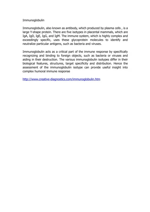
Introduction • 1 of 5 classes of antibodies (IgG, IgM, IgA, IgDand IgE) • 0.002% of serum antibodies • Half-life = 2 days • Fc portion binds to mast cells and basophils where is mediates many allergic reactions. • Fc portion binds to Eosinophils enabling opsonization against parasitic infections.
Causes of Hyper IgE • Most common cause of elevated IgE • Western countries – allergies • Third world – parasitic infections. • Extreme levels (800 – 25,000IU/ml) • Severe atopic dermatitis • ABPA • Parasitic infections • IgE myeloma • Buckley Syndrome (Job’s Syndrome or Hyper IgE syndrome)
HIES • Identified in 1966 by Davis Schaller • Two girls w/ red hair, chronic dermatitis and recurrent staphylococcus abscesses • Disease was named after the Biblical Job
Hyper IgE Syndrome (HIES) • Autosomal dominant or recessive • Dominant – pts fail to lose primary teeth so can have two sets of teeth simultaneously • Recessive – severe viral infections and neurologic sequelae, often fatal in childhood • Characteristics: • Frequent Staphylococcal skin infections • Eczema-like skin rash • Severe lung infections – pneumatoceles • Very high levels of IgE (>2000 IU/ml)
Pathophysiology • Abnormal neutrophil chemotaxis (↓interferon µ) is postulated as cause of disease. • This defect proved inconsistently present. • IgE usually >10x normal • ↑ Eos are common in 90%
Diagnosis • Clinical diagnosis • Immune defects • Somatic defects • Elevated IgE • NIH developed a scoring system • Not for clinical use • Linkage studies to determine inheritance patterns.
Treatment • Chronic antibiotics – given repeated staph infections • Diagnosis should ideally be made in childhood to prevent pneumatocele formation • Good skin care • I&D of abscesses • Mucocutaneous candidiasis the most frequent co-infection.
Treatment • Typically pts are unaware of how sick they are • Fever and other markers of inflammation may not be present. • Empyema is frequently present and requires drainage. • Pulmonary cavities are at high risk for co or super-infection. • Extensive bronchiectasis.
Treatment • Immunomodulators have been unsuccessful • Levamisole – only RTC • INF-µ -- was inconsistent in its effects on IgE levels and infections. • Cyclosporine A – used successfully in Israel but results not officially published. • IVIG – reasonable option given that encapsulated organisms are most often at fault.
References • GrimbacherBodo, Steven M Holland, Puck Jennifer, Hyper-IgE syndromes. Immunological Reviews 2005. Vol 203: 244-250 • http://www.clinlabnavigator.com/Test-Interpretations/immunoglobulin-e-ige.html • Para FM – Extreme increase of total IgE with Eosinophilia, case report; AllergolImmunolClin 2000;15:194-197.