Download
1 / 122
1.27k likes | 1.57k Views
4 – Lab The Tissue Level of Organization. An Introduction to Tissues. Learning Outcomes 4-1 Identify the four major types of tissues in the body and describe their roles. 4-2 Discuss the types and functions of epithelial tissue.

E N D
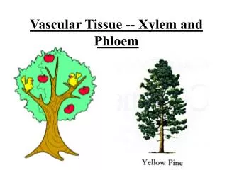
4 – Lab The Tissue Level of Organization
An Introduction to Tissues • Learning Outcomes • 4-1 Identify the four major types of tissues in the body and describe their roles. • 4-2 Discuss the types and functions of epithelial tissue. • 4-3 Describe the relationship between form and function for each type of epithelium.
An Introduction to Tissues • Learning Outcomes • 4-4 Compare the structures and functions of the various types of connective tissues. • 4-5 Describe how cartilage and bone function as a supporting connective tissue. • 4-6 Explain how epithelial and connective tissues combine to form four types of tissue membranes, and specify the functions of each. • 4-7 Describe how connective tissue establishes the framework of the body.
An Introduction to Tissues • Learning Outcomes • 4-8 Describe the three types of muscle tissue and the special structural features of each type. • 4-9 Discuss the basic structure and role of neural tissue • 4-10 Describe how injuries affect the tissues of the body. • 4-11 Describe how aging affects the tissues of the body.
An Introduction to Tissues Tissues Structures with discrete structural and functional properties Tissues in combination form organs, such as the heart or liver Organs can be grouped into 11 organ systems
4-1 Four Types of Tissue • Tissue • Are collections of cells and cell products that perform specific, limited functions • Four types of tissue • Epithelial tissue • Connective tissue • Muscle tissue • Neural tissue
4-1 Four Types of Tissue • Epithelial Tissue • Covers exposed surfaces • Lines internal passageways • Forms glands • Connective Tissue • Fills internal spaces • Supports other tissues • Transports materials • Stores energy
4-1 Four Types of Tissue • Muscle Tissue • Specialized for contraction • Skeletal muscle, heart muscle, and walls of hollow organs • Neural Tissue • Carries electrical signals from one part of the body to another
4-2 Epithelial Tissue • Epithelia • Layers of cells covering internal or external surfaces • Glands • Structures that produce secretions
4-2 Epithelial Tissue • Characteristics of Epithelia • Cellularity (cell junctions) • Polarity (apical and basal surfaces) • Attachment (basement membrane or basal lamina) • Avascularity • Regeneration
Figure 4-1 The Polarity of Epithelial Cells Cilia Microvilli Apicalsurface Golgiapparatus Nucleus Mitochondria Basement membrane Basolateralsurfaces
4-2 Epithelial Tissue • Functions of Epithelial Tissue • Provide Physical Protection • Control Permeability • Provide Sensation • Produce Specialized Secretions (glandular epithelium)
4-3 Classification of Epithelia • Singular = Epithelium; Plural = Epithelia • Classes of Epithelia • Based on shape • Squamous epithelia — thin and flat • Cuboidal epithelia — square shaped • Columnar epithelia — tall, slender rectangles • Based on layers • Simple epithelium — single layer of cells • Stratified epithelium — several layers of cells
4-3 Classification of Epithelia • Squamous Epithelia • Simple squamous epithelium • Absorption and diffusion • Mesothelium • Lines body cavities • Endothelium • Lines heart and blood vessels
Figure 4-3a Squamous Epithelia Simple Squamous Epithelium LOCATIONS: Mesothelia lining ventral body cavities; endothelia lining heartand blood vessels; portions of kidney tubules (thin sections of nephron loops); inner lining of cornea; alveoli of lungs FUNCTIONS: Reduces friction; controls vessel permeability; performsabsorption and secretion Cytoplasm Nucleus Connective tissue LM 238 Lining of peritoneal cavity
4-3 Classification of Epithelia • Squamous Epithelia • Stratified squamous epithelium • Protects against attacks • Keratin protein adds strength and water resistance
Figure 4-3b Squamous Epithelia Stratified Squamous Epithelium LOCATIONS: Surface of skin; lining of mouth, throat, esophagus, rectum, anus, and vagina FUNCTIONS: Provides physical protection against abrasion, pathogens, and chemical attack Squamoussuperficial cells Stem cells Basementmembrane Connectivetissue Surface of tongue LM 310
4-3 Classification of Epithelia • Cuboidal Epithelia • Simple cuboidal epithelium • Secretion and absorption • Stratified cuboidal epithelia • Sweat ducts and mammary ducts
Figure 4-4a Cuboidal and Transitional Epithelia Simple Cuboidal Epithelium LOCATIONS: Glands; ducts;portions of kidney tubules; thyroidgland Connectivetissue FUNCTIONS: Limited protection,secretion, absorption Nucleus Cuboidalcells Basementmembrane Kidney tubule LM 650
Figure 4-4b Cuboidal and Transitional Epithelia Stratified Cuboidal Epithelium LOCATIONS: Lining of some ducts(rare) FUNCTIONS: Protection, secretion,absorption Lumenof duct Stratifiedcuboidalcells Basementmembrane Nuclei Connectivetissue Sweat gland duct LM 500
4-3 Classification of Epithelia • Transitional Epithelium • Tolerates repeated cycles of stretching and recoiling and returns to its previous shape without damage • Appearance changes as stretching occurs • Situated in regions of the urinary system (e.g., urinary bladder)
Figure 4-4c Cuboidal and Transitional Epithelia Transitional Epithelium LOCATIONS: Urinarybladder; renal pelvis;ureters FUNCTIONS: Permitsexpansion and recoilafter stretching Epithelium(relaxed) Basement membrane Connective tissue andsmooth muscle layers LM 400 Empty bladder Epithelium(stretched) Basement membrane LM 400 Connective tissue andsmooth muscle layers LM 400 Full bladder Urinary bladder
4-3 Classification of Epithelia • Columnar Epithelia • Simple columnar epithelium • Absorption and secretion • Pseudostratified columnar epithelium • Cilia movement • Stratified columnar epithelium • Protection
Figure 4-5a Columnar Epithelia Simple Columnar Epithelium LOCATIONS: Lining ofstomach, intestine, gallbladder,uterine tubes, and collectingducts of kidneys Microvilli Cytoplasm FUNCTIONS: Protection,secretion, absorption Nucleus Basementmembrane Looseconnective tissue LM 350 Intestinal lining
Figure 4-5b Columnar Epithelia Pseudostratified Ciliated Columnar Epithelium LOCATIONS: Lining ofnasal cavity, trachea, andbronchi; portions of malereproductive tract Cilia Cytoplasm FUNCTIONS: Protection,secretion, move mucuswith cilia Nuclei Basementmembrane Looseconnective tissue Trachea LM 350
Figure 4-5c Columnar Epithelia Stratified Columnar Epithelium LOCATIONS: Small areas ofthe pharynx, epiglottis, anus,mammary glands, salivarygland ducts, and urethra Looseconnective tissue Deeper basalcells FUNCTION: Protection Superficialcolumnar cells Lumen Lumen Cytoplasm Nuclei Basementmembrane Salivary gland duct LM 175
4-3 Classification of Epithelia • Glandular Epithelia • Endocrine glands • Release hormones • Into interstitial fluid • No ducts • Exocrine glands • Produce secretions • Onto epithelial surfaces • Through ducts
4-3 Classification of Epithelia • Glandular Epithelia • Gland Structure • Unicellularglands • Mucous (goblet) cells are the only unicellular exocrine glands • Scattered among epithelia • For example, in intestinal lining
4-3 Classification of Epithelia • Gland Structure • Multicellular glands • Structure of the duct • Simple (undivided) • Compound (divided) • Shape of secretory portion of the gland • Tubular (tube shaped) • Alveolar or acinar (blind pockets) • Relationship between ducts and glandular areas • Branched (several secretory areas sharing one duct)
Figure 4-7 A Structural Classification of Exocrine Glands Duct SIMPLE GLANDS Glandcells SIMPLETUBULAR SIMPLE COILEDTUBULAR SIMPLE BRANCHEDTUBULAR Examples: Examples: Examples: • Gastric glands • Merocrine sweat • Intestinal glands • Mucous glands glands of esophagus,tongue, duodenum SIMPLE ALVEOLAR(ACINAR) SIMPLE BRANCHEDALVEOLAR Examples: Examples: • Not found in adult; a • Sebaceous (oil) stage in developmentof simple branchedglands glands
Figure 4-7 A Structural Classification of Exocrine Glands COMPOUND GLANDS COMPOUND TUBULOALVEOLAR COMPOUNDTUBULAR COMPOUND ALVEOLAR(ACINAR) Examples: Examples: Examples: • Mucous glands (in mouth) • Salivary glands • Mammary glands • Bulbo-urethral glands (in • Glands of respiratory male reproductive system) passages • Testes (seminiferous • Pancreas tubules)
4-4 Connective Tissue • Characteristics of Connective Tissue • Specialized cells • Solid extracellular protein fibers • Fluid extracellular ground substance • The Extracellular Components of Connective Tissue (Fibers and Ground Substance) • Make up the matrix • Majority of tissue volume • Determinesspecialized function
4-4 Connective Tissue • Functions of Connective Tissue • Establishing a structural framework for the body • Transporting fluids and dissolved materials • Protecting delicate organs • Supporting, surrounding, and interconnecting other types of tissue • Storing energy reserves, especially in the form of triglycerides • Defending the body from invading microorganisms
4-4 Connective Tissue • Classification of Connective Tissues • Connective tissue proper • Connect and protect • Fluid connective tissues • Transport • Supporting connective tissues • Structural strength
4-4 Connective Tissue • Categories of Connective Tissue Proper • Loose connective tissue • More ground substance, fewer fibers • For example, fat (adipose tissue) • Dense connective tissue • More fibers, less ground substance • For example,tendons
Fibroblasts Fibrocytes Adipocytes Mesenchymal cells Macrophages Mast cells Lymphocytes Microphages Melanocytes 4-4 Connective Tissue Connective Tissue Proper Cell Populations
4-4 Connective Tissue • Fibroblasts • The most abundant cell type • Found in all connective tissue proper • Secrete proteins and hyaluronan (cellular cement) • Fibrocytes • The second most abundant cell type • Found in all connective tissue proper • Maintain the fibers of connective tissue proper
4-4 Connective Tissue • Adipocytes • Fat cells • Each cell stores a single, large fat droplet • Mesenchymal Cells • Stem cells that respond to injury or infection • Differentiate into fibroblasts, macrophages, etc.
4-4 Connective Tissue • Macrophages • Large, amoeba-like cells of the immune system • Eat pathogens and damaged cells • Fixed macrophages stay in tissue • Free macrophages migrate
4-4 Connective Tissue • Mast Cells • Stimulate inflammation after injury or infection • Release histamine and heparin • Basophils are leukocytes (white blood cells) that also contain histamine and heparin
4-4 Connective Tissue • Lymphocytes • Specialized immune cells in lymphatic (lymphoid) system • For example, lymphocytes may develop into plasma cells (plasmocytes) that produce antibodies
4-4 Connective Tissue • Microphages • Phagocytic blood cells • Respond to signals from macrophages and mast cells • For example,neutrophils and eosinophils • Melanocytes • Synthesize and store the brown pigment melanin