Download

1 / 83
850 likes | 1.14k Views
Localized Prostate Cancer. Epidemiology. Incidence : - Prostate cancer is the fourth most common male malignant neoplasm worldwide . - Worldwide, consistent increase in its incidence .

E N D
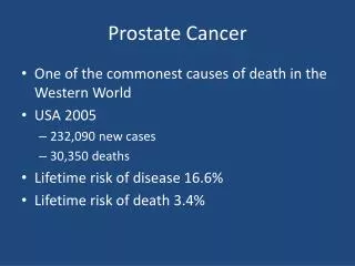
Epidemiology Incidence: - Prostate cancer is the fourth most common male malignant neoplasm worldwide . - Worldwide, consistent increase in its incidence . - Prostate cancer has been the most common visceral malignant neoplasm in U.S. men since 1984 . - lifetime risk of disease : - 17.6% for whites - 20.6% for African Americans . - lifetime risk of death : - 2.8% for whites - 4.7% for African Americans . - The risk of developing clinical cancer is 10% .
Racial Differences & nationality - black men have the highest incidence of prostate cancer - 170 per 100,000 U.S black men - 110 per 100,000 U.S. white men) - overall incidence of prostate cancer lower in Asians, Hispanics, and Natives (70 per 100,000) . - Scandinavian countries have particularly high rate of prostate ca . - Asian countries have lowest prostate cancer incidence . - Environment plays an important role in modulating prostate cancer risk around the world .
The incidence in Saudi Arabia - 2,270 Saudi men older than 50 years screened from 1994 to 1997 . - The incidence of prostatic carcinoma in the Kingdom of Saudi Arabia is low(3.1/100,000 person-years ) despite a high saturated fat diet in recent years (Hanash KA et al,2000) .
Age at Diagnosis : - Prostate cancer is rarely diagnosed in men younger than 50 years,(<0.1%). - Peak incidence occurs between the ages of 70 and 74 years, with 85% diagnosed after the age of 65 years
Mortality -The 3nd most common cause of cancer death . - The mortality rate for prostate cancer in white men in the United States has declined to a level lower than that observed before the introduction of PSA-based screening in 1987 ( Tarone et al, 2000 ) . -Postmortem : 10% 3rd decade 34% 5th decade 67% 9th decade
Risk Factors: - Age: - Microscopic foci in 30% of men in their 50s and 70% of men over the age of 80 . - Rare below 40y, increase with age. - 29% 6th decade - 67% 9th decade - Geographic Variation: Western nations more than the Asian & Far East . - Race: Blacks more than white. - Family History.
Hereditary Prostate Cancer (HPC) - 10%of prostate cancer - 45% in patients < 55years old - Younger < 60y - Chromosome 1q ,8p, Xp ,mutation BRCA2 gene - 1st degree relative One -------------- 2 fold increased risk Two --------------- 5 fold Three ---------------- 11 fold - Sporadic & familial prostate cancers behave similarly
Promoters (+ve): - Androgen - Genetic polymorphism -Insulin-like growth factor 1 (IGF-1) - Fatty diet - Calcium
Protective (-ve) - Lycopene. - Selenium. - Vitamin E. - Vitamin D.
What about ??? Vasectomy………….. Controversial !!! Sexual activity X Smoking X Height & weight. X Alcohol consumption x
Etiology Normal prostate epithelium Prostate cancer
PathologyPrecursor lesions to invasive carcinoma ● Prostatic intraepithelial neoplasia (PIN) ● Atypical small acinar proliferation (ASAP) Both are reported by pathologists as “ suspicious for cancer”
PIN - Benign prostatic acini & ducts lined by atypical cells - B.M intact, focally fragmented - Low-grade PIN 1 , high grade PIN2 & PIN3 - High grade PIN precursor for high grade prostate cancer
HGPIN - Its finding in peripheral zone biopsy carries 30-40% prediction for cancer prostate at a subsequent biopsy . - site of PIN is not an indicative of the subsequent cancer site, nor always in a cancer containing prostate . Cancer can arise without PIN
Atypical small acinar proliferation - Acini lined by cytologically abnormal epithelial cells. - Columnar cells have prominent nucleoli. - B.M intact, basal layer focally absent Cancer in subsequent biopsy 40% .
Pathology Type: - 95%Adenocarcinoma - Mucinous adenocarcinoma, small cell carcinoma - Transitional , mesenchymal Location: 75% peripheral zone 20% transitional zone 5% central zone 85% multifocal
Spread 1-local - Extrprostatic extension Tumor >0.5 cm - preineural invasion - S.V invasion - Rectum 2-Lymphatic Tumor >4cm PSA>20ng/ml 3-Metastasis Bone PSA>10ng/ml Lung, bladder, liver ,adrenal gland & testis .
Grading Gleason system 1 ---5 (gland architecture at low magnification) Multifocal pattern Two grades ( most dominant+ 2nd dominant) To give a sum 2 -10 Factors affect the Gleason's: Hormone therapy BPH 5 alph reductase inhibitors
Grading 2 – 4 well differentiated 5 – 7 moderately differentiated 8 – 10 poorly differentiated => 7 poor prognosis Correlates with prognosis Ex. 3+3=6 worse prognosis than 3+2=5 Dominant grade correlates with prognosis Ex. 4+3=7 worse prognosis than 3+4=7
Diagnosis ◙ History : symptoms ◙ Physicalexamination, DRE ◙ PSA ◙ TRUS ◙ Biopsy
Clinical Presentation Locally Advanced: - Asymptomatic . - LUTS. - Haematospermia. - Haematuria. - Perineal discomfort - Symptoms of renal faliure - with ureteric obstruction - Malignant priapasm (rare) - Rectal obstruction (rare) Localized cancer: -Asymptomatic( incidental PSA, DRE) screening - LUTS - Haematospermia - Haematuria - Perineal discomfort.
Clinical Presentation: Metastatic disease: - Asymptomatic with occult disease. (PSA, DRE) - Lower limb swelling ,lymphatic obstruction. - Anorexia, weight loss. - Bone pain, pathological fracture. - Neurological (spinal cord compression) - Anaemia. - Dyspnoea, jaundice, bleeding tendency .
DRE : Abnormal DRE: - Asymmetry, nodule ,fixed craggy mass . - 50% abnormal DRE associated with cancer prostate . - 40% of DRE diagnosed cancers are organ confined .
PSA : - 34KD glycoprotein ,human kallikrein (hK3) . - Columnar acinar & ductal prostatic epithelial cells . - 75% protein bound , 25% free . - Half-life 2.2days +/- 0.8 . - High positive predictive value for cancer.
PSA Prostate specific not cancer specific. - Cancer prostate. - BPH - UTI. - Acute prostatitis. - Chronic prostatitis - Retention . - Catheterization, biopsy, TURP. - DRE, ejaculation. PSA level vary with age, race and prostatic volume Finastride 12 months , 50% reduction in PSA .
:PSA When to check serum PSA? 1- Screening: 2- Suspected prostate cancer: - LUTS - Abnormal DRE - Progressive bone pain - Unexplained anaemia, wt loss, anorexia - Spontaneous thrombo-embolism . 3- Monitoring cancer prostate patients
PSA • Normal < 4 ng/ml ?? > 2.5 - 4 -10 ng/ml cancer - PSA>10ng/ml - >50% patients has extra prostatic disease. - 20% L.N +ve - PSA>50ng/ml - 66% L.N involvement - 90% seminal vesicle involvement - 20% cancer prostate with normal PSA .
PSA ◙To increase the specificity of PSA ● Age- adjusted normal range for PSA ● F:T PSA ratio. ● PSA density. ● PSA velocity
Age –adjusted normal PSA ● 40 -49 y 0 – 2.5ng/ml ● 50 –59y 0 – 3.5ng/ml ● 60 -69y 0 – 4.5ng/ml ● 70 -79y 0 – 6.5ng/ml
PSA Density ●PSA 4 – 10 ng/ml ● PSA / Prostate volume ● < 0.15 /mg PSA Velocity ● PSA increase/year ● > 0.75 ng/ml per year suggest cancer.
Free to total PSA - Free PSA <25% prognostic factor for prostate cancer. - 10% or less associated with 50% incidence of cancer prostate. - Ex. -Man with normal DRE, PSA 4-10ngml . - 60% F:T 10% -Has (27%) risk of cancer :-- -10% F:T >25%
TRUS - Outline the prostate - Prostate volume. • Hypo- hyperechoic lesions • - Cysts, abscess, calcifications - Prostatic lesions, biopsy
TRUS - Tumor size : Not successful - S.V involvement: - In S.V invasion, 90% of them has an abnormal sonogram. - 15 -40% normal appearing S.V contains cancer.
Prostatic Biopsy Trans rectal, perineal Indications:- - Abnormal DRE . - Elevated PSA . - Previous biopsies showed PIN or ASAP - Previous normal biopsy but PSA is rising or DRE become abnormal . - To confirm viable prostate cancer following treatment if further treatment will be considered .
Prostatic Biopsy Protocol: • Antimicrobial coverage pre &post - 6 – 12 tru-cut needle biopsies (18G) Complications: - Vaso-vagal fainting - 0.5% septicemia. - 0.5% rectal beeding - Mild haemospermia or haematuria for 3 weeks .
Prostatic Biopsy Sextant biopsies: - Lateral directed biopsies are better than direct biopsies . - False negative 15 -34% . If patient is still suspicious for cancer & biopsy is negative Repeat biopsy 8-12 core biopsies
Staging ■ - T - DRE, PSA, TRUS, Biopsy ■ - N - imaging +/- FNA ■ - M - bone scan, CT, MRI - molecular staging
CT scan • Inadequate for local tumor staging . -cannot image cancer within the prostate . - insufficient resolution for minimal ECE . • Insufficient sensitivity and specificity for LN status • Limited value for local recurrence in PSA faliure . - Seletive indications in high risk pt with PSA>20, T3, high Gleason grade .
MRI • Better than CT scan on local lesion. • Body MRI :- role in SV extension, not for ECE . • Endorectal coli MRI:- - better to delineate location, size and extent of cancer within prostate and location of ECE . • Indicated in moderate and high risk patient . • Accuracy :- 50 --- 90% . • Dis :- cost, time, invasiveness .
Radionucleide Bone scan -Standerd staging tool . -Indications:- - high risk disease - PSA > 10 in newly diagnosed . - PSA > 20 during follow up . - high grade & stage . - suggestive signe or symptoms .