Download
1 / 21
210 likes | 389 Views
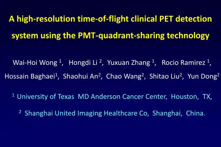
A high-resolution time-of-flight clinical PET detection system using the PMT-quadrant-sharing technology. Wai -Hoi Wong 1 , Hongdi Li 2 , Yuxuan Zhang 1 , Rocio Ramirez 1 , Hossain Baghaei 1 , Shaohui An 2 , Chao Wang 2 , Shitao Liu 2 , Yun Dong 2

E N D
A high-resolution time-of-flight clinical PET detection system using the PMT-quadrant-sharing technology Wai-Hoi Wong 1, Hongdi Li 2, Yuxuan Zhang 1, Rocio Ramirez 1, Hossain Baghaei1, ShaohuiAn2, Chao Wang2, ShitaoLiu2, Yun Dong2 1 University of Texas MD Anderson Cancer Center, Houston, TX, 2 Shanghai United Imaging Healthcare Co, Shanghai, China.
System Design • LYSO is expensive (1/5 the price of gold) • The Objective is to get higher sensitivity per cc of LYSO • More efficient use of LYSO by increasing the axial field of view • while reducing the crystal depth • GEANT4 MC simulation • But large AFOV increases • PMT and electronics cost • Use PMT-quadrant-sharing • reducing PMT usage by 75% • for increasing AFOV cheaply • 15mm deep LYSO, 28-cm AFOV
Detector Ring Design • To achieve ultrahigh resolution using large PMT to increase AFOV 2.35 x 2.35 mm pitch 38-mm PMT 16 x 16 LYSO block Achieve decoding 256 crystals per PMT usage • Adapt PMT-Quad-Sharing blocks to a gapless detector-ring geometry Ring has 24 modules (3 x 7 blocks) 3 blocks in-plane and 7 blocks axial The edge blocks are half-ground to fit the quadrant-sharing PMT
A detector module 27.6-cm axial FOV
The “Slab-Sandwich-Slice” (SSS) Detector Production There are 4 sandwich types in this 16 x 16 block Each reflecting mask can be cut into any shape providing many degrees of freedom to optimize crystal decoding These are 15 sets of inter-slab irregular reflecting masks for a 16 x 16 array to decode 256 crystals / PMT
The SSS production method is highly uniform and precise as shown in the decoding map of these 72 detector blocks
We use the 5th-generation HYPER Pileup-event-recovery front-end electronics Hybrid coincidence
NEMA image resolution measurement • Reconstruction algorithm: FBP2D (SSRB) • Pixel Size: 0.3 x 0.3 x 1.22
PSF reconstructed image resolution (mm) V: Vertical H: Horizontal
Very fine axial sampling, slice-to-slice separation = 1.22 mm
Oncology 3 minutes/bed 4 bed positions
TOF + PSF 2 min/bed, 2 iterations PSF 3 min/bed, 3 iterations MIP TOF + PSF Recon SNR: TOF/PSF 2 min/bed 2 iterations = PSF 3 min/bed 3 iterations 2/3 scan time, faster recon, no loss of detectability SNR = (Signal – Background) / SDBackground See Jakoby, et al, Phys. Med. Biol. 56
Conclusions • With PMT-quadrant-sharing Detector design we have developed an ultrahigh resolution TOF PET/CT • It has a resolution of 2.8 mm using FBP (1.5 mm using PSF) • Large axial FOV 27.6 cm, ultrafine axial sampling of 1.22 mm • This large system with ultrahigh resolution uses only 576 PMT (reducing PMT and electronics cost, while increasing reliability) • It has 129,024 detectors with a TOF resolution of 473 ps
These developments have been supported by: • NIH-RO1- EB001038 PHS Grant • NIH-RO1- EB001481 PHS Grant • NIH-RO1- EB004840 PHS Grant • Shanghai United Imaging Healthcare Fund