Download
1 / 1
10 likes | 65 Views
Analysis of ASF distribution in neuronal nuclei during neurofibrillary degeneration stages. ASF presence, loss, and effects on fibrillar material observed. Immunostained sections reveal ASF localization and NFT morphology.

E N D
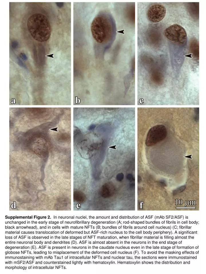
Supplemental Figure 2. In neuronal nuclei, the amount and distribution of ASF (mAb SF2/ASF) is unchanged in the early stage of neurofibrillary degeneration (A; rod-shaped bundles of fibrils in cell body; black arrowhead), and in cells with mature NFTs (B; bundles of fibrils around cell nucleus) (C; fibrillar material causes translocation of deformed but ASF-rich nucleus to the cell body periphery). A significant loss of ASF is observed in the late stages of NFT maturation, when fibrillar material is filling almost the entire neuronal body and dendrites (D). ASF is almost absent in the neurons in the end stage of degeneration (E). ASF is present in neurons in the caudate nucleus even in the late stage of formation of globose NFTs, leading to misplacement of the deformed cell nucleus (F). To avoid the masking effects of immunostaining with mAb Tau1 of intracellular NFTs and nuclear tau, the sections were immunostained with mSF2/ASF and counterstained lightly with hematoxylin. Hematoxylin shows the distribution and morphology of intracellular NFTs.