Download
1 / 25
270 likes | 594 Views
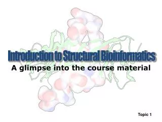
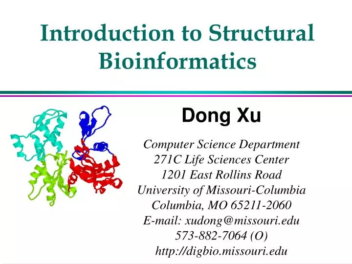
Introduction to Structural Bioinformatics. Dong Xu Computer Science Department 271C Life Sciences Center 1201 East Rollins Road University of Missouri-Columbia Columbia, MO 65211-2060 E-mail: xudong@missouri.edu 573-882-7064 (O) http://digbio.missouri.edu. Structural Bioinformatics.

E N D
Introduction to Structural Bioinformatics Dong Xu Computer Science Department 271C Life Sciences Center 1201 East Rollins Road University of Missouri-Columbia Columbia, MO 65211-2060 E-mail: xudong@missouri.edu 573-882-7064 (O) http://digbio.missouri.edu
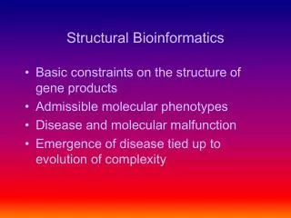
Structural Bioinformatics • Prediction and modeling • Protein structure • DNA structure • RNA structure • Membrane structures • Large-complex structure
An Overview • A protein folds into a unique 3D structure under the physiological condition Lysozyme sequence (129 amino acids): KVFGRCELAA AMKRHGLDNY RGYSLGNWVC AAKFESNFNT QATNRNTDGS TDYGILQINS RWWCNDGRTP GSRNLCNIPC SALLSSDITA SVNCAKKIVS DGNGMNAWVA WRNRCKGTDV QAWIRGCRL Protein backbones: Side chain
Protein Structure Representations Lysozyme structure: ball & stick strand surface
Growth of Protein Data Bank (PDB) [ PDB: http://www.pdb.org ]
Protein Structure Database: PDB (1) • PDB (Protein Data Bank) Web site: http://www.rcsb.org/pdb/ • 33,252 Structures as of 25-Oct-2005 • PDB ID: 4-character identifier (1cau, 1gox, and 256b) • Search methods * search by PDB ID (e.g. 1lyz); * SearchLite: protein name, author's name, etc. (e.g., HIV protease); * SearchFields: EC Number, the name of the binding ligand (e.g., inhibitor), the range of the protein size, and the secondary structure content.
Protein Structure Database: PDB (2) PDB format (headers + coordinates): HEADER OXIDOREDUCTASE (OXYGEN(A)) 14-JUN-89 1GOX 1GOX COMPND GLYCOLATE OXIDASE (E.C.1.1.3.1) 1GOX ... ATOM 232 N ALA 29 54.035 64.332 19.352 1.00 23.93 1GOX ATOM 233 CA ALA 29 52.992 65.356 19.569 1.00 24.74 1GOX ATOM 234 C ALA 29 53.519 66.762 19.309 1.00 25.43 1GOX ATOM 235 O ALA 29 54.648 67.179 19.655 1.00 25.66 1GOX ATOM 236 CB ALA 29 52.433 65.340 20.993 1.00 24.54 1GOX ... HETATM 3165 O HOH 658 62.480 62.480 0.000 0.50 65.79 1GOX ... END
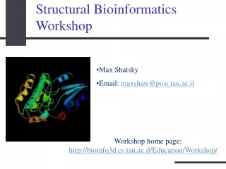
Molecular Visualization RasMol: http://www.umass.edu/microbio/rasmol/index2.htm VMD: http://www.ks.uiuc.edu/Research/vmd
Relevance of Protein Structurein the Post-Genome Era structure medicine sequence function
Structure-Function Relationship Certain level of function can be found without structure. But a structure is a key to understand the detailed mechanism. A predicted structure is a powerful tool for function inference. Trp repressor as a function switch
Structure-Based Drug Design Structure-based rational drug design is still a major method for drug discovery. HIV protease inhibitor
Structures in Protein Language: Letters Words Sentences Protein: Residues Secondary Structure Tertiary Structure
a helix • Single protein chain (local) • Shape maintained byintramolecular H bondingbetween -C=O and H-N-
b sheet • Several protein chains • Shape maintained byintramolecular H bondingbetween chains • Non-local on protein sequence
Protein Structure Domain (1) • Structure domain: compact, globular unit glycoprotein actin
Protein Structure Domain (2) • Structure domain is evolutionary, functional, and folding unit of a protein • Domain insertion: insert: zinc metalloproteinase + parent: thioredoxin (disulfide oxidoreductase) Dsba: disulfide bond forming protein • Protein design (growth hormone) • Threading target
Structure Is Better Conserved during Evolution Structure can adopt a wide range of mutations. Physical forces favor certain structures. Concept of fold. Number of fold is limited. Currently ~800 Total: 1,000s ~10,000s TIM barrel
The number of different protein folds is limited Already known folds PDB submissions per year New folds Year
Protein Folding Problem A protein folds into a unique 3D structure under the physiological condition Lysozyme sequence: KVFGRCELAA AMKRHGLDNY RGYSLGNWVC AAKFESNFNT QATNRNTDGS TDYGILQINS RWWCNDGRTP GSRNLCNIPC SALLSSDITA SVNCAKKIVS DGNGMNAWVA WRNRCKGTDV QAWIRGCRL
Web Addresses • Resource: http://digbio.missouri.edu/resource/ • Further reading (a review on protein modeling): www.bentham.org/cpps1-1/Dong%20Xu/xu_cpps.htm