Download
1 / 26
310 likes | 685 Views
Anatomy of the Eye. Mr. Young Anatomy & Physiology. YOUR EYES . . . 70% of ALL SENSORY RECEPTORS in the body 1,000,000 nerve fibers per eye more than 2 million working parts average person blinks 12 times per minute - about 10,000 blinks in an average day

E N D
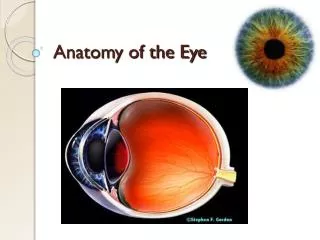
Anatomy of the Eye Mr. Young Anatomy & Physiology
YOUR EYES . . . • 70% of ALL SENSORY RECEPTORS in the body • 1,000,000 nerve fibers per eye • more than 2 million working parts • average person blinks 12 times per minute - about 10,000 blinks in an average day • can distinguish 500 shades of the gray. • contribute towards 85% of your total knowledge. • always the same size from birth
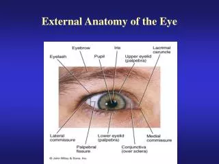
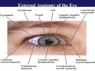
Accessory Structures • Eyelids • Conjunctiva • Lacrimal apparatus • SECRETIONS: • Sebum (oil): tarsal glands • Mucus: conjunctiva • Tears: lacrimal gland • Antibodies • Lysozymes
Accessory Structures • Extrinsic muscles
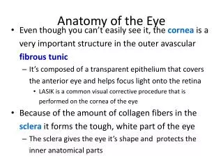
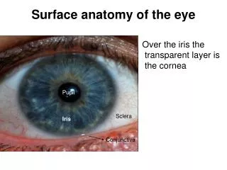
The Fibrous Tunic • Sclera • White connective tissue layer • Seen anteriorly as the “white of the eye” • Cornea • Transparent, central anterior portion • Allows for light to pass through • Repairs itself easily • The only human tissue that can be transplanted without fear of rejection
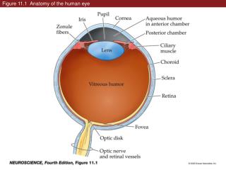
Choroid Layer • Blood-rich nutritive tunic • Pigment prevents light from scattering • Modified interiorly into two structures • Cilliary body – smooth muscle • Iris • Pigmented layer that gives eye color • Pupil – rounded opening in the iris
Sensory Tunic (Retina) • Contains receptor cells (photoreceptors) • Rods • Cones • Signals pass from photoreceptors via a two-neuron chain • Bipolar neurons • Ganglion cells • Signals leave the retina toward the brain through the optic nerve
Neurons of the Retina and Vision • Rods • Most are found towards the edges of the retina • Allow dim light vision and peripheral vision • Perception is all in gray tones
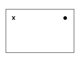
Neurons of the Retina and Vision • Cones • Allow for detailed color vision • Densest in the center of the retina • Fovea centralis – area of the retina with only cones • No photoreceptor cells are at the optic disk, or blind spot
Cone Sensitivity • There are three types of cones • Different cones are sensitive to different wavelengths • Color blindness is the result of lack of one cone type Figure 8.6
What about non-visual light perception? • Video 1 • Video 2
Astigmatism • Unequal curvatures in lens/cornea
Now . . . • . . . a few tests to reinforce the role of the brain in vision. • http://www.eyecanlearn.com/