Download
1 / 48
620 likes | 984 Views
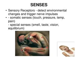
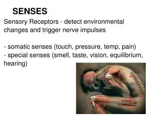
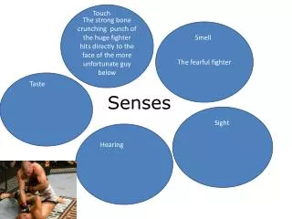
Senses. Chapter 18. Sensory receptors. First structure in a reflex arc Specialized for specific stimuli May be encapsulated or bare neuron endings Exterioreceptors detect stimuli outside body Ex) light, sound, Indirectly involved in homeostasis Interioreceptors

E N D
Senses Chapter 18
Sensory receptors • First structure in a reflex arc • Specialized for specific stimuli • May be encapsulated or bare neuron endings • Exterioreceptors • detect stimuli outside body • Ex) light, sound, • Indirectly involved in homeostasis • Interioreceptors • detect stimuli inside body • Ex) blood pressure, body temperature, water balance • Directly involved in homeostasis
Sensory receptors cont’d. • Chemoreceptors • Taste buds, olfactory, pain, receptors in aortic and carotid bodies monitoring blood pH • Photoreceptors • Rods • black and white vision • cones • color vision • Mechanoreceptors • Auditory, touch, equilibrium, pressoreceptors for blood pressure and stretch of lung tissue • Thermoreceptors • body temperature
Exteroreceptors • Table 18.1
Sensation • Occurs when nerve impulses arrive at cerebral cortex • Perception • when the cerebral cortex interprets meaning of the impulses • Impulses conveyed to the cord and then through ascending tracts to brain • Sensation we perceive depends on where impulses originate • All impulses are in the form of action potentials • For example: • If action potentials travel to visual cortex a visual image is perceived • If they travel to the auditory cortex a sound is heard
Sensation • Fig 18.1
Sensations cont’d. • Integration • Occurs before receptors initiate action potentials • Summing up of signals • Sensory adaptation – a type of integration • Decrease in response to a stimulus • May occur because receptors stop responding to a repeated stimulus • May occur because the thalamus screens out repeated impulses from a specific area • Sensory receptors make a significant contribution to homeostasis
Proprioreceptors • Mechanoreceptors in muscles and tendons • Gives us an unconscious “body sense” • Awareness of position of limbs • Degree of muscle and tendon stretch • Equilibrium and posture • Example: Muscle spindles • Wrapped around muscle fibers, monitor degree of stretch and initiate reflexes to adjust • Example: knee jerk reflex
Muscle spindle • Fig 18.2
Cutaneous Receptors • In the dermis of the skin • Allow skin to be sensitive to touch, pressure, and temperature • Fine touch receptors • Meissner corpuscles-fingertips, lips, palms, penis, clitoris • Merkel disks- junction of epidermis and dermis • Root hair plexus- free nerve endings at base of follicles • Allows sensation when hair is touched
Cutaneous Receptors cont’d • Pressure receptors • Pacinian corpuscles: onion-shaped, deep in dermis • Ruffini endings and Krause end bulbs: encapsulated receptors with complex nerve networks • Pain receptors • Free nerve endings • Damaged tissues release chemicals that stimulate pain receptors
Cutaneous Receptors cont’d • Fig 18.3
Cutaneous receptors cont’d. • Referred pain • In some areas stimulation of internal pain receptors is also perceived as pain from the skin • We believe impulses from internal pain receptors also synapse in cord with neurons receiving pain impulses from skin • Ex: pain originating in heart is also referred to left arm and shoulder
Taste • Receptors in tongue, hard palate, pharynx, epiglottis • Specific regions of the tongue are sensitive to particular tastes • Tip- sweet; Margins- salty and sour; back –bitter • Taste bud structure • Each has a pore surrounded by supporting cells and taste cells • Taste cells have microvilli with receptors • How the brain receives taste information • Chemicals bind to receptors on microvilli-impulses generated • Gustatory (taste) cortex surveys incoming pattern of impulses • “Weighted average” is the perceived taste
Taste buds • Fig 18.4
Smell • 80-90% of what we perceive as taste is actually smell • Olfactory receptor structure • Located high in nasal cavity on olfactory membrane • Olfactory cells have a tuft of olfactory cilia with receptors • Each olfactory cell has receptors of only 1 type • Inhaled molecules bind to receptors to generate impulses • How the brain receives odor information • Nerve fibers lead to olfactory bulb-extension of brain, also linked to limbic system-odors can trigger emotions and memories • Single odor composed of many different molecules-activates a characteristic combination of receptor proteins • Odor’s “signature” is interpreted by brain
Olfactory cell location and anatomy • Fig 18.5
Vision: Eye Structure • Sclera- outer layer • White fibrous covering except for corneal region • Cornea- transparent collagen fibers • Choroid- middle layer • Thin, darkly pigmented, vascular • Absorbs stray light rays • Includes iris at front • controls pupil size • Iris is pigmented to give the eye it’s color • Behind the iris the choroid thickens to form the ciliary body • Ciliary body controls curvature of the lens
Vision: Eye Structure cont’d. • Choroid layer cont’d. • Lens • attached to the ciliary body by suspensory ligaments • divides the inner eyeball into chambers • Anterior chamber- between the cornea and the lens • Filled with water aqueous humor • Small amount produced each day while same amount is drained through small ducts • Glaucoma- build up of fluid when ducts are blocked; can cause increased intraocular pressure and blindness • Posterior chamber- between lens and back of eyeball • Filled with gelatinous vitreous humor
Vision: Eye Structure cont’d. • Retina- innermost layer of eyeball • Lines the posterior compartment • Contains the photoreceptors • Rods- function in black and white dim-light vision • Evenly distributed throughout retina • Cones- function in bright-light color vision • Concentrated in fovea centralis • Sensory fibers leave the retina to form the optic nerve
Vision: Eye Structure cont’d. • Fig 18.6
Vision: Eye Functions • Lens • Focuses light rays onto the retina • Image is inverted and upside down on the retina • If eyeball is too long or too short corrective lenses are needed to bring image into focus on the retina
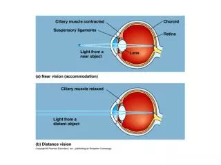
Vision: Eye Functions cont’d. • Lens cont’d • Visual accommodation • For viewing close objects • Ciliary muscle contracts which releases tension on suspensory ligaments • Lens rounds up due to natural elasticity • Increases refraction (bending) of light rays to bring them into focus on the lens • Presbyopia-age changes decrease elasticity of lens • Distance Vision • Ciliary muscle relaxes • Increases tension on the suspensory ligaments • Causes lens to flatten out • This brings light rays from distant objects into focus on the retina
Focusing • Fig 18.7
Vision: Eye Functions cont’d. • Photoreceptors • General structure • Outer segment joined to inner segment by a stalk • Pigment located in disks within outer segments • Synaptic vesicles located at synaptic endings of inner segments • Rods • Visual pigment is rhodopsin • Complex molecule made up of the protein opsin and retinal which is a derivative of vitamin A • When rod absorbs light rhodopsin is split to opsin + retinal • Closes ion channels in rod cell membrane • Stops release of inhibitory transmitter signals • Signals sent to other neurons in retina and on to optic nerve
Photoreceptors in the eye • Fig 18.8
Vision: Eye Functions cont’d. • Photoreceptors cont’d • Cones • 3 kinds of cones each with a specific pigment • B (blue), G (green), and R (red) pigments • Each is an iodopsin composed of retinal and an opsin • Opsin is slightly different in each • Combinations of cones are simulated by in-between colors • Mechanics of light absorption and stimulus generation is the same as in rods • Cones allow color vision which is of higher acuity than the rods
Vision: Eye Functions cont’d. • Retina • 3 layers of cells • Layer closest to choroid contains rods and cones • Middle layer composed of bipolar cells • Inner layer composed of gangion cells • Rod and cone cells synapse with bipolar cells which synapse with ganglion cells- axons of these cells become the optic nerve • Many more photoreceptors than ganglion cells • 150 rods stimulate a single ganglion cells • 1 cone stimulates 1 ganglion cell • Explains why color vision is higher acuity
Vision: Eye Functions cont’d. • Function of retina cont’d. • Integration occurs as signals pass to bipolar and ganglion cells • Each ganglion cell receives signals from about 1 square mm of retina • This region is the ganglion cell’s receptive field • Ganglion cell is stimulated only by signals received from the center of its receptive field • Otherwise it is inhibited • If all rod cells in the receptive field receive light the cell responds in a neutral way-reacts weekly or not at all • Considerable processing occurs in the retina before ganglion cells generate impulses • Impulses from the ganglion cells travel in the optic nerve to the visual cortex where further integration occurs
Structure and function of the retina • Fig 18.9
Vision: Eye Functions cont’d. • Blind spot • Area on retina where optic neurons leave to form the optic nerve • There are no photoreceptors here!
Vision: Perception • Optic nerves from each eye travel to the optic chiasma • Some of the axons cross over at the optic chiasma • Fibers from the right half of each retina join together to form the right optic tract • Fibers from the left half of each retina join together to form the left optic tract • Optic tracts travel around the hypothalamus and most fibers synapse with nuclei in the thalamus • Axons from the thalamic nuclei form optic radiations that carry impulses to the visual cortex on each side • Right and left visual cortex must communicate for us to see entire visual field
Optic chiasma • Fig 18.10
Vision: Abnormalities • Color blindness • Complete colorblindness is rare • Most common types involve deficiency in one type of cone • Red-green colorblindess • Most common type • X-linked recessive trait • 5-8% of the male population
Vision: Abnormalities cont’d. • Myopia • Nearsighted • Can see close objects better than distant ones • Eyeball is elongated so image is brought to point focus in front of the retina • Corrected by concave lenses which diverge light rays so point focus is farther back • Farsighted • Can see distant objects better than close ones • Eyeball is shortened so image is brought to point focus behind the lens • Corrected by convex lenses to increase bending of light rays so point focus is farther forward
Common abnormalities of the eye, with possible corrective lenses • Fig 18.11
Hearing: Ear Anatomy • Outer ear • Pinna • Auditory canal • Middle ear • Tympanic membrane-beginning of the middle ear • Auditory ossicles (bones) • Malleus, incus, stapes • Forms a “bridge” across the middle ear • Auditory tube-extends from middle ear to nasopharynx • Helps equalize pressure across the tympanic membrane • Oval window and round window- membranous connections with inner ear • Inner ear • Semicircular canals, vestibule, and cochlea
Hearing: Ear Anatomy cont’d • Fig 18.12
Hearing: Auditory Pathway • Sound waves channeled into the auditory canal by the pinna • Strike the tympanic membrane and it begins to vibrate • Vibrations are amplified across the ossicles • Amplified about 20 times • Stapes is attached to oval window- • Oval window vibrates and transmits vibrations to fluid inside the cochlea
Hearing: Auditory Pathway cont’d. • Cochlear structure • 3 canals • Vestibular canal • Cochlear canal • Tympanic canal • Spiral organ (organ of Corti) is located in the cochlear canal • Consists of hair cells and a gelatinous tectorial membrane • Hair cells sit on the basilar membrane and have stereocilia embedded in the tectorial membrane
Hearing: Auditory Pathway cont’d. • Stapes causes oval window to vibrate • Vibrations move from vestibular canal to tympanic canal across the basilar membrane • Basilar membrane moves up and down and the stereocilia of the hair cells bend • Generates nerve impulses in the cochlear nerve • Travel to the brain • Auditory cortex interprets them as sound
Hearing: Auditory Pathway cont’d. • Each part of the spiral organ is sensitive to different wave frequencies or pitch of sound • Tip = low pitches, base = high pitches • Nerve fibers along length lead to slightly different areas of auditory cortex • Pitch we hear depends on which region of the basilar membrane is vibrating and which area of the auditory cortex is stimulated • Volume is a function of amplitude of sound waves • Loud sounds cause greater vibrations of basilar membrane • Increased stimulation is interpreted as volume • Brain interprets tone based on distribution of hair cells stimulated
Functions of the parts of the ear • Table 18.3
Ear: Sense of Equilibrium • 3 semicircular canals each processing different motions • Horizontal motion, Head tilting, Rotation • Rotational Equilibrium Pathways • Receptors are found in the ampulla of the semicircular canals and contain hair cells with stereocillia. • Hair cells in the ampulla have cilia embedded in a gel-like mass, the cupula. • As fluid within a canal flows and bends a cupula, the stereocilia are bent and this changes the pattern of impulses carried in the vestibular nerve to cerebellum and cerebrum • Brain uses this information to make postural corrections • Vertigo- dizziness and sense of rotation • Motion sickness- from continuous movement of fluid in semicircular canals
Equilibrium cont’d. • Gravitational equilibrium pathway • Depends on utricle and saccule • Utricle is sensitive to horizontal movements of the head • Saccule is sensitive to vertical movements of the head • Both contain hair cells with stereocilia are embedded in otolithic membrane • Large central cilium called the kinetocilium • Calcium carbonate granules (otoliths) rest on otolithic membrane • When head or body moves in horizontal or vertical plane the otoliths are displaced and the otolithic membrane sags
Sense of equilibrium cont’d. • Gravitational equilibrium pathway cont’d. • If stereocilia are bent move toward the kinetocilium, nerve impulses increase in the vestibular nerve • If sterocilia are bent away from the kinetocilium, nerve impulses decrease in the vestibular nerve • When a person is upside down, impulses in the vestibular nerve stop • Vestibular cortex uses this information to determine movement of the head • Initiates appropriate motor output to right the body’s current position in space
Ear: Sense of Equilibrium cont’d • Fig 18.14