Download
1 / 47
500 likes | 760 Views
Thoracic and Lumbar Spine Fractures and Dislocations: Assessment and Classification. Mark L Prasarn MD University of Texas Dept of Orthopaedic Surgery Houston, Texas Updated 7/2016. Anatomy of the Spine. Cervical. Five segments or divisions C/T/L/S/C Made up of: 7 cervical 12 thoracic

E N D
Thoracic and Lumbar Spine Fractures and Dislocations: Assessment and Classification Mark L Prasarn MD University of Texas Dept of Orthopaedic Surgery Houston, TexasUpdated 7/2016
Anatomy of the Spine Cervical • Five segments or divisions • C/T/L/S/C • Made up of: • 7 cervical • 12 thoracic • 5 lumbar • 5 sacral • 4 coccygeal • 4 curves in the sagittal plane • T 20-40º • L 50º Thoracic Lumbar Sacral Coccygeal
Anatomy of Thoracic Spine • Kyphosis 20-40° • Facet orientation coronal • Ribs and sternum provide stable “ring” • Narrow canal • Cord injury • Rigid, with little motion
Anatomy of the Lumbar Spine • Lordosis ~50° • Facet orientation sagittal • Very mobile • Large vertebral bodies • Cauda equina
Thoracolumbar Junction • Common site of injury • Most between T11 to L2 • Transitional anatomy • Rigid T spine • Flexible L spine • CoronalSagittal facets • KyphoticLordotic spine
Thoracolumbar Junction • Altered biomechanics • Natural curves able to absorb and dissipate axial loads in subjacent regions • Straight T-L junction allows less shock absorption • Cannot disperse force
Patient Evaluation • Pre-hospital care • EMT personnel • Initial assessment • Transport and immobilization • Spinal precautions have reduced complete injuries and improved survival!
Patient Evaluation • ABC’s of Trauma • History • Physical Examination • Neurological Classification
Physical Examination Ecchymosis where posterior ligaments disurpted, gap and step-off on exam • Inspection • Palpation • Sensory Exam • Motor Exam • Reflex Evaluation • Including Bulbocavernosus
21 y/o female MVC ejection • Seat belt sign • Severe tenderness and gap between SP at T11-T12 • Flexion distraction injury*
Neurologic Injury • Exam must include spinal cord function as well as nerve root and peripheral n. integrity • Cord terminates at L1 • Injuries can cause variable picture: • Cord injury • Conus medullaris • Cauda Equina • Root injury
Neurological Injury • Radiculopathy • Dermatomal sensory changes • Myotomal weakness • Hyporeflexia • More diffuse injury possible cord/conus/cauda injury • If potential for cord injury must check BC reflex Actual cord transection from fracture
Incomplete vs. Complete • Bulbocavernosis reflex • Return heralds end of spinal shock • Must check perianal sensation and rectal tone • Presence of sacral sparing is the key factor! • Critical for prognosis
Complete versus Incomplete • ASIA A is complete • ASIA B has sacral sparing only • ASIA C is 3/5 strength in <50% of the muscle groups below the neurological level • ASIA D is 3/5 strength in >50% of the muscle groups below the neurological level • ASIA E is full strength and sensation
Incomplete Patterns • Central cord – motor groups in upper extremities more affected than lower extremities • Anterior cord – motor loss with some sensory preservation, vibration and proprioception intact • Posterior cord – motor preservation with loss of proprioception and vibratory sense • Brown-Sequard – ipsilateral motor, vibration, and proprioception loss with contralateral sensory loss • Conus medullaris – pain with isolated loss of bowel/bladder, usually between T12 and L1
Spinal Cord Injury - Prognosis • Complete SCI – studies show will not walk again • Incomplete Syndromes • Central Cord - UE>LE • Brown-Sequard - Good Prognosis • Anterior Cord - Worst Prognosis • Posterior Cord – Rare • Cauda Equina Syndrome
Radiology - Morophology • Plain films • Lateral • Wedge-shaped • Loss of ant body height • Post elements? • Kyphosis • Anteroposterior • Interpedicular widening • Height loss • CT scan • Canal compromise • Comminution • Facets Empty facet sign
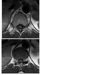
Utility of MRI • MRI – images spinal cord, intervertebral discs, ligamentous structures, rule out epidural hematoma, also incorporated into some classification systems
Spinal Stability? • “the loss of the ability of the spine under physiologic loads to maintain its pattern of displacement so that there is no initial or additional neurological deficit, no major deformity, and no incapacitating pain” White & Panjabi 1990
Instability • Any dislocation • Spondyloptosis • Facet dislocation • Rotational/Translational injury • Compression/Burst Fxs • Area of much controversy • Still no clear determinant • Look at Posterior ligamentous complex (PLC)
Instability • Palpable gap or step-off • Progressive neurological deficit • Progressive kyphosis • Radiographic significant posterior column injury • >50% height loss? • Yuan et al. Spine 1982 • MRI? (PLC key!)
MRI to determine Instability • Lee et al. Spine 2000 • 34 patients • MRI compared with operative findings • 30 w/ MRI findings • Significant correlation
Classifications Necessary for…… • Uniform method of description • Directing treatment *** • Facilitating outcome analysis • Should be: Comprehensive Reproducible Usable Accurate
1 2 3 Anatomic Classification3 Column TheoryDenis 83 • Based on radiographic review of 412 cases • 5 types, 20 subtypes • Anterior- ALL , anterior 2/3 body • Middle - post 1/3 body, PLL • Posterior- all structures posterior to PLL • Same as Holdsworth • Posterior injury-not sufficient to cause instability
AO Mechanistic Classification • Review of 1445 cases (Magerl, Gertzbein et al. European Spine Journal 1994) • Based on direction of injury force • 3 types,53 injury patterns • Type A - Compression • Type B - Distraction • Type C - Rotational Increasing severity
Thoracolumbar Injury Classification and Severity Score TLICS Vaccaro et al. JSDT 2005 • Developed by STSG • 3 major variables: • MOI • Integrity of post ligs • Neurologic status • Quantified by severity score • 1-4 for each category • Reflects severity of injury and contribution on stability
MOI based on Radiographs Most severe level for multi-level injuries 3 Categories: Compression Translation/Rotation Distraction Compression Simple 1 Burst 2 Compression + >15 coronal deformity 2 Trans/Rotation 3 Distraction 4 TLICS Vaccaro et al. JSDT 2005
Neurologic Status Intact 0 Root 2 Complete 2 Incomplete or Cauda Equina 3 Posterior Ligament Complex High correlation to MRI (Lee et al. Spine 2000) Fat suppressed T2 Score None 0 Indeterminate 2 Definite 3 TLICS Vaccaro et al. JSDT 2005
TLICS Vaccaro et al. JSDT 2005 • Treatment • ≤3 Non-op • ≥5 Operative • 4 Either • Must consider other “clinical qualifiers” • Kyphosis, collapse, sternal fx, CHI, polytrauma, AS, DISH, Obesity, Med Comorbidities, etc.
Burst Fxs • Substantial axial load • Compression failure of ant + mid columns • Falls & MVCs • Most have some canal compromise
Stable Burst Fxs • No posterior ligamentous disruption • Cantor et al. Spine 1993 • 18 pts tx’d in TLSO • No progression of deformity • No neuro deficit • Resorption observed
Most do not require surgery! • Key is PLC! • Some consider: • Kyphosis >25° • >50% height loss • >50% canal compromise Controversial***
Operative If deficit and lamina fx, then likely dural tear Cammisa JBJS 1989
Surgical treatment • Anterior • Posterior • Both • Percutaneous • Fusion or no fusion • Short or long construct Percutaneous stabilization of multiple fractures
Load Sharing Classification (McCormack SPINE 94) • Devised method of predicting posterior failure or short segment failure • 1-3 points assigned to the variables below • Sum the points for a 3-9 scale • <6 points posterior only • >6 points anterior <3° 4-9° 0-1mm 1-2mm >2mm <30% 30-60% >10° >60%
Folman and Gepstein, J Orthop Trauma, 2003 • 85 pts reviewed to determine late outcome of non-op management • Chronic pain predominant in 69.4% • 25% of subjects had changed jobs (most full to part) • 48% of subjects filed lawsuits concerning injury • Pain intensity correlated with angle of kyphosis • But not w/magnitude of anterior column deformity • Bed rest alone adequately manages traumatic, uncomplicated thoracolumbar wedge fractures
Dai, J Trauma, 2004 • 147 pts w/acute thoracolumbar fractures: 1988 to 1997 • Min. 3yr f/u; 4 pts died during hospital stay • Delayed diagnosis in 28 pts (19%) • Differences b/w surgical & non: • in pulmonary complications & length of hospital stay in non-op pts. • Surgical pts had highly significantly less pain • Radiographic studies should be performed • Choice of treatment in pts with multiple injuries is not different from that in pts with no asscd injuries
Risk Factors for Respiratory Failure Following OperativeStabilization of Thoracic and Lumbar Spine FracturesTimothy P. McHenry, Sohail K. Mirza, JingJing Wang, Charles E. Wade, Grant E. O'Keefe, Andrew T. Dailey, Martin A. Schreiber and Jens R. Chapman. J Bone Joint Surg Am. 2006;88:997-1005 • January 1985 through January 2004 • Trauma registry compared to ARDS registry • 140 / 1032 operative thoracolumbar fractures developed respiratory failure
Risk Factors for Respiratory Failure Following Operative Stabilization of Thoracic and Lumbar Spine Fractures • Surgical timing is the only modifiable risk factor • Surgical stabilization before 48 hours may reduce the development of pulmonary failure
Conclusions on Treatment • Surgically treating incomplete neuro deficits potentiates improvement and rehabilitation • Complete neuro deficits may benefit from operative treatment to allow mobilization • Little chance of developing neuro deficits with nonoperative treatment
Thank You Selected Images from: Court-Brown, C. et al. Rockwood & Greens Fractures in Adults. Philadelphia: Lippincott Williams & Wilkins, 2014