Download
1 / 31
310 likes | 340 Views
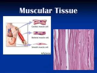
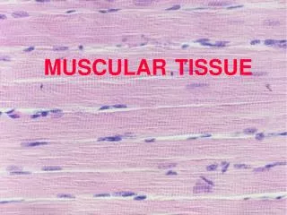
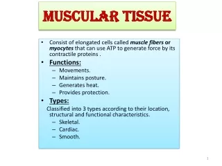
Muscular Tissue. Types of Muscle Tissue. skeletal cardiac smooth. Skeletal. attached to bones, skin, deep fascia, or other muscles voluntary control striated , alternating light and dark bands along length of myofibrils many nuclei Functions: - movement - posture - respiration.

E N D
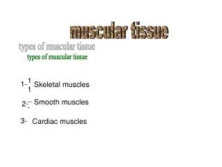
Types of Muscle Tissue • skeletal • cardiac • smooth
Skeletal • attached to bones, skin, deep fascia, or other muscles • voluntary control • striated , alternating light and dark bands along length of myofibrils • many nuclei • Functions: - movement - posture - respiration
Skeletal Muscle Nuclei Striation
Cardiac • located only in the heart • striated, single nucleus, branched fibers with intercalated discs • involuntary control by autonomic nervous system • regulation of heart rate is primarily due to hormones and neurotransmitters • no regeneration capability • propels blood through blood vessels
Cardiac Muscle intercalated disc
Smooth • located in hollow organs, skin attached to hair follicles, etc. • no striations, single nucleus, spindle-shaped fibers • involuntary control by autonomic nervous system • some regeneration
Functions of Smooth Muscles • mix and propel food though GI tract • regulate flow of blood by changing diameter of lumen • contraction of urinary bladder, gallbladder, and spleen, expels urine, bile and blood • control sphincter muscles • control muscles of eye • contraction of arrector pili muscles
Multiunit Smooth • muscle fibers are not well organized • occur as separate fibers rather than sheets • found in irises of eye, walls of blood vessels
Visceral Smooth • composed of sheets of spindle-shaped cells • in contact with one another • more common type • found in hollow visceral organs • capable of stimulating each other • display rhythmicity due to self-exciting fibers - responsible for peristalsis
Peristalsis • wavelike motion • occurs in various tubular organs • helps force contents of these organs along their lengths
Contraction of Smooth Muscles • acetylcholine and norepinephrine • also affected by hormones • slower to contract - slower to relax • can maintain a forceful contraction longer than skeletal with same amount of ATP • can change length without changing tautness
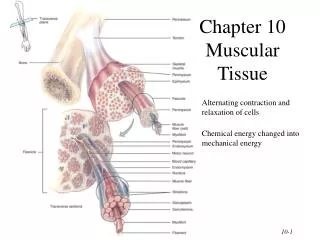
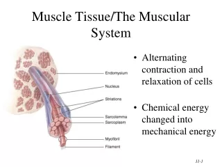
Muscle Fibers • many muscle fibers are enclosed in a delicate connective tissue sheath called endomysium • several sheathed fibers are wrapped in perimysium in bundles called fascicles (10 -100 fibers)
Muscle Fibers (cont.) • many fascicles are joined together by even tougher covering called epimysium • fascia covers entire muscles which lead into tendons which attach to bones
Individual Muscle Fiber (single cell) • sarcolemma - plasma membrane covering of muscle cell • sarcoplasm - cytoplasm of a skeletal muscle cell
Individual Muscle Fiber (cont.) • sarcoplasmic reticulum - network of membranous channels - within sarcoplasm (corresponds to endoplasmic reticulum) - surrounds each myofibril - channels run parallel to myofibril - stores calcium which is necessary for muscle contraction
Individual Muscle Fiber (cont.) • transverse tubules - fingerlike inward invaginations or channels of sarcolemma - extend from membrane and pass through the fiber - open to outside of the muscle fiber - contain extracellular fluid - carry action potentials to sarcoplasmic reticulum
Individual Muscle Fiber (cont.) • cisternae - enlarged portions of sarcoplasmic reticulum - lie on either side of transverse tubules - near region where actin and myosin overlap
Individual Muscle Fiber (cont.) • myofibrils - long ribbon-like organelles - lie parallel to one another • myofilaments - thread-like structures within myofibrils (contain two types of protein filaments) • actin (thin & light) and myocin (thick & dark)
Actin and Myosin • appear as light (thin) and dark (thick) bands • arrangement of these fibers produces the characteristic striations of a skeletal muscle fiber • slide past each other causing muscle cells to contract
Myosin • located within the dark portions of the striations (A bands)
Actin • located primarily within light areas (I bands) • during muscle contraction actin filaments slide farther into A bands • attached to the Z lines at end of I bands • Z lines extend across muscle fiber enabling adjacent myofibrils to lie side by side • segment between two Z lines is called a sarcomere
Sarcomeres • repeating units composed of filaments inside myofibrils • do not extend the entire length of the muscle fiber
Characteristics of Muscle Tissue • excitability • contractility • extensibility • elasticity
Excitability(irritability) • ability to respond to stimuli • generate action potentials or impulses • stimuli that initiate action potentials in muscles are neurotransmitters • neurotransmitters are released by axon terminals of neurons
Contractility • ability to contract and shorten to generate a force • muscles contract in response to action potentials
Extensibility • ability to be stretched or extended when pulled • with pairs of skeletal muscles - one muscle is contracted while the opposing one is usually stretched
Elasticity • ability to return to original shape after contraction or extension