Download
1 / 27
280 likes | 314 Views
Learn about the structure, functions, and distribution of major human immunoglobulins (Ig) in the humoral basis of immunity. Explore the classes of Ig, antibody diversity, and abnormal Ig in physiological and pathological contexts.

E N D
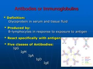
Immunoglobulins • Humoral basis of immunity – 19th century. Introduction of an Ag into an animal Abs appeared in the serum & body fluids Immune sera • Abs react with Ag in a specific & observable manner.
Distribution of Major Human Ig • Electrophoresis of human serum separated serum proteins into 2 major components : 1. Soluble Albumins 2. Insoluble Globulins - , & • Ab activity - γ globulin fraction of serum proteins. • 1964 – ‘Immunoglobulin’ by WHO.
Igs are synthesised mainly by the plasma cells & to some extent by the lymphocytes. • Constitutes 20-25% of total serum proteins. • 5 classes of Igs – IgG, IgA, IgM, IgD & IgE.
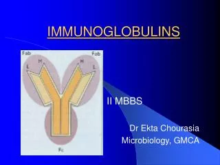
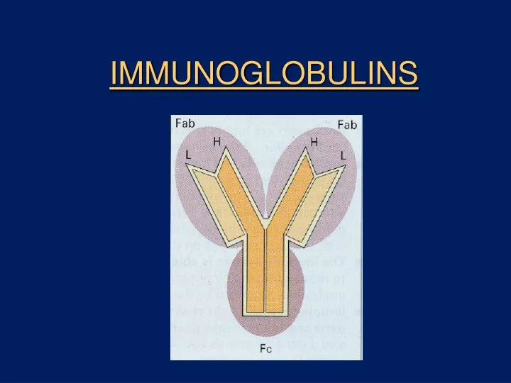
Structure of an Ig • Glycoproteins. • 2 pairs of polypeptide chains – 2 light (L) chains & 2 heavy (H) chains. • “L” chain – smaller chain - low molecular wt (25,000) • “H” chain – larger chain - high molecular wt (50,000)
Structure • L chain attached to H chain by disulphide & non- covalent bonds. • L chains : 2 forms – kappa () & lambda () • Each molecule of Ig can have either or , but never both. • H chains : structurally & antigenically distinct for each class.
H chain • H chain designated by Greek letter.
Structure • Light chain – 2 regions Constant (CL ) Variable (VL ) C – terminal N - terminal
Structure • H chain also divided into VH & CH regions; the CH region is further divided into CH1, CH2 & CH3. • Regions also called as DOMAINS : - globular in shape - stabilized by intrachain disulphide bonds • Ag binding sites are located in the variable domains.
Hypervariable regions • Amino acid sequence in the variable region of L & H chains are not uniformly variable. • Consists of some highly variable(hypervariable) & some relatively invariable zones. • Highly variable zones actually make contact with the epitope on an Ag and are called as Complementarity Determining Regions (CDRs) • 3 CDRs – each made up of 9 -12 amino acids. CDR3 is the longest & most variable of the three.
Structure • ‘ Hinge ’ region – segment of H chain between CH1 & CH2. • Flexibility to Ab • Susceptible to enzymes & chemicals. • Studies involving the cleavage of Ig molecule by pepsin & papain have led to a detailed picture of Ig structure.
Fragments of Ig • Fab – Ag binding. Fc fragment • Composed of carboxy terminal of H chain. • Determines the biological properties of Ig molecule. • Receptors for Fc portion expressed by – mononuclear cells neutrophils phagocytosis NK cells tumour cell killing eosinophils & mast cell degranulation mast cells
Ig G • Major serum Ig • Constitutes 75% of total Igs. • 4 subclasses found in humans – IgG1, IgG2, IgG3 & IgG4, each having a distinct type of gamma chain. • Major Ab of secondary response, found both in serum & body fluids. • Only maternal Ig to be transported across placenta – natural passive immunity in newborn. • Participates in complement fixation, precipitation & neutralisation of viruses & toxins.
Ig M • 5-8 % of serum Igs. • Short lived Abs. • Pentameric structure. • Predominant Ab in primary immune response. • Earliest Ab to be synthesized by the fetus. • Confined to the intravascular pool due to its large size. • Not transported across placenta. • Presence of IgM in newborn indicates intra uterine infection. • Useful in the diagnosis of congenital infections like syphilis, rubella, HIV, toxoplasmosis etc.
Ig A • 2nd most abundant, constitutes 10-13 %. • Major Ig in the colostrum, saliva, tears & other body fluids. • Occur in 2 forms : IgA1 & IgA2. • Secretory IgA is always in dimeric form – composed of 2 basic chain units, a J chain & the secretory component. • Secretory component helps to transport the dimer from the submucosa to the mucosal cell surface. • Secretory component protects IgA from proteolytic digestion and denaturation.
Ig D • Resembles Ig G structurally. • Occurs along with Ig M on the surface of B cells • Very susceptible to proteolytic attack.
Ig E • Present in very low levels in serum. • Found on the surface of mast cells & basophils which have specific receptors for the Fc portion of IgE. • Chiefly produced in the linings of respiratory & intestinal tracts. • Responsible for anaphylactic type of hypersensitivity. • Defense against parasitic infections.
Abnormal Igs • Structurally similar proteins in serum seen in certain pathological conditions. • Bence Jones protein in multiple myeloma – light chains of Igs. • Cryoglobulinemia – formation of gel or ppt on cooling the serum which redissolves on warming – in myelomas, SLE etc.
Antibody diversity • An individual produces a large number of Abs to cope with the vast number of different Ags. • This Ab diversity is due to the Ig genes. • Genes coding for the variable & constant portions of the chains are separate • One or only few genes code for C region whereas many genes code for the V region.
Generation of diversity • Multiple V- region genes. • V-J & V-D-J recombination. • Junctional diversity • Nucleotide addition – extra nucleotides may get inserted between VH & D, and between D & JH segments • Somatic mutation – point mutation in the genes for V domain.