Download
1 / 67
680 likes | 999 Views
Congenital Heart Lesions. Dominic Blurton MD PCA Pediatric Cardiology. Outline. Normal anatomy 1.L -> R shunt 2.Left side obstruction 3.Cyanotic heart lesions Right side obstruction and R -> L shunt Transposition 4.Mixing Lesions Surgical therapy. Ductus Arteriosus. Aorta.

E N D
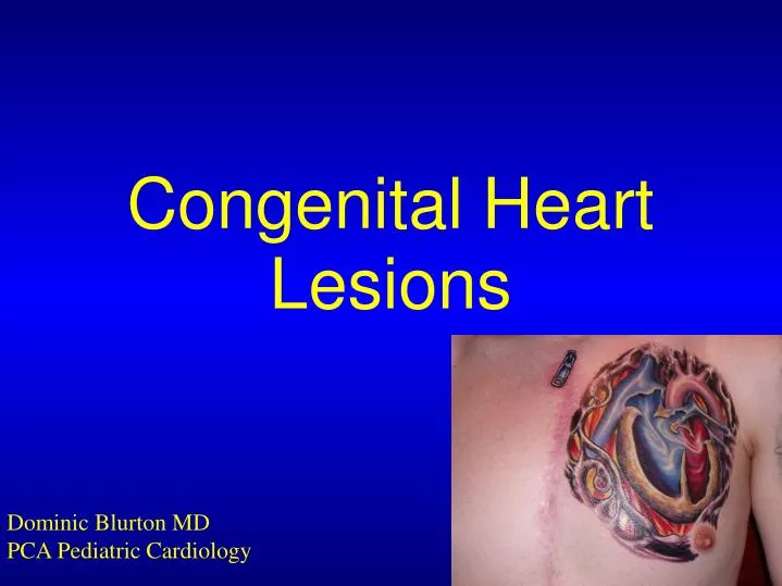
Congenital Heart Lesions Dominic Blurton MD PCA Pediatric Cardiology
Outline Normal anatomy 1.L -> R shunt 2.Left side obstruction 3.Cyanotic heart lesions Right side obstruction and R -> L shunt Transposition 4.Mixing Lesions Surgical therapy
Ductus Arteriosus Aorta Pulmonary Artery Left Atrium Patent Foramen Ovale Right Atrium LeftVentricle Right Ventricle
Key Points Blood flows to the path of least resistance Pulmonary resistance < systemic resistance All newborns have connections PDA PFO
Physiological classification of defects • 1.L -> R shunt • 2.Left side obstruction • 3.Cyanotic heart lesions • Right side obstruction and R -> L shunt • Transposition • 4.Mixing Lesions
Outline Normal anatomy L -> R shunt Left side obstruction Cyanotic heart lesions Right side obstruction and R -> L shunt Transposition Mixing Lesions Surgical therapy
Left to right shunting Right and left side connected Increased (too much) pulmonary blood flow Respiratory distress/ CHF
Left to right shunt lesions Ventricular septal defect (VSD) Atrial septal defect (ASD) AV canal Patent ductus arteriosus (PDA)
Outline Normal anatomy L -> R shunt Left side obstruction Cyanotic heart lesions Right side obstruction and R -> L shunt Transposition Mixing Lesions Surgical therapy
Left side obstruction Not enough blood to the body Hypo-perfusion, acidosis, shock
Left side obstructive lesions Mitral valve obstruction Aortic valve obstruction Coarctation of the aorta Everything obstructed Hypoplastic left heart syndrome
Outline Normal anatomy L -> R shunt Left side obstruction Cyanotic heart lesions Right side obstruction & R -> L shunt Transposition Mixing Lesions Surgical therapy
Cyanotic lesions Connection - right and left sides AND right side obstruction Decreased pulmonary blood flow OR Separated systems
Cyanotic lesions Right side obstructions Tricuspid obstruction Pulmonary obstruction Tetralogy of Fallot Separate systems Transposition of the great vessels
Outline Normal anatomy L -> R shunt Left side obstruction Cyanotic heart lesions Right side obstruction & R -> L shunt Transposition Mixing Lesions Surgical therapy
Mixing lesions Very large intra or extracardiac connection Key points- What goes into the lungs comes out of the lungs = red What goes into the body comes out of the body = blue May have right side obstruction
Mixing Lesions Single ventricle Double inlet left ventricle (DILV) Double outlet right ventricle (DORV) Primitive ventricle Hypoplastic right or left ventricle Total anomalous pulmonary venous return (TAPVR) Truncus arteriosus
Outline Normal anatomy L -> R shunt Left side obstruction Cyanotic heart lesions Right side obstruction & R -> L shunt Transposition Mixing Lesions Surgical therapy
Surgical therapy Repair vs. palliation Palliating a single ventricle - Example: HLHS Stage I: Norwood and BT shunt Stage II: Glenn shunt Stage III: Fontan