Download
1 / 48
480 likes | 774 Views
The Cellular Environment: Fluids and Electrolytes, Acids and Bases. Chapter 3. Distribution of Body Fluids. Total body water (TBW) 60% of total body weight Intracellular fluid – inside the cells Extracellular fluid – not encased in cells

E N D
The Cellular Environment: Fluids and Electrolytes, Acids and Bases Chapter 3
Distribution of Body Fluids • Total body water (TBW) 60% of total body weight • Intracellular fluid – inside the cells • Extracellular fluid – not encased in cells • Interstitial fluid – found in between cells and tissues • Intravascular fluid- plasma found in circulatory system • Lymph, synovial, intestinal, biliary, hepatic, pancreatic, CSF, sweat, urine, pleural, peritoneal, pericardial, and intraocular fluids are extracellular
Water Movement Between the ICF and ECF • Osmolality – the concentrations of solutes in water • Osmotic forces – solutes will influence the movement of water across membranes • Aquaporins- water channel proteins in membranes • Starling hypothesis • Net filtration = forces favoring filtration – forces opposing filtration • As fluid flows through capillary it looses water and create greater osmotic return of water as it flows toward veinule end of capillary
Net Filtration • Forces favoring filtration • Capillary hydrostatic pressure (blood pressure) • Interstitial oncotic pressure (water-pulling) • Forces favoring reabsorption • Plasma oncotic pressure (water-pulling) • Interstitial hydrostatic pressure
Edema • Accumulation of fluid within the interstitial spaces • Causes: • Increase in hydrostatic pressure • Losses or diminished production of plasma albumin • Increases in capillary permeability • Lymph obstruction – elephantitus, flibitus
Water Balance • Thirst perception • Osmolality receptors in medula respond to osmotic pressue of ECF • Hyperosmolality and plasma volume depletion • ADH secretion from posterior pituitary – conserves water in kidney to maintain water balance
Sodium and Chloride Balance • Sodium • Primary ECF cation • Regulates osmotic forces • Roles • Neuromuscular irritability, acid-base balance, and cellular reactions • Chloride • Primary ECF anion • Provides electroneutrality
Sodium and Chloride Balance • Renin-angiotensin system – substanced produced in both liver and kidney • Angiotensin produced by liver and coverted by enzymes activated by renin from Kidney Juxta Glomerular Aparatus to a powerful vasoconstrictor. • Aldosterone – hormone from adrenal gland to regulate Na and K • Natriuretic peptides • Atrial natriuretic peptide - hormone from heart • Brain natriuretic peptide – hormone from brain • Urodilantin (kidney) – Kidney hormone
Alterations in Na+, Cl–, and Water Balance • Isotonic alterations • Total body water change with proportional electrolyte and water change • Isotonic volume depletion • Isotonic volume excess
Hypertonic Alterations • Hypernatremia • Serum sodium >147 mEq/L • Related to sodium gain or water loss • Water movement from the ICF to the ECF • Intracellular dehydration • Manifestations • Intracellular dehydration, convulsions, pulmonary edema, hypotension, tachycardia, etc.
Water Deficit • Dehydration • Pure water deficits • Renal free water clearance • Manifestations • Tachycardia, weak pulses, and postural hypotension • Elevated hematocrit and serum sodium level
Hypochloremia • Occurs with hypernatremia or a bicarbonate deficit • Usually secondary to pathophysiologic processes • Managed by treating underlying disorders
Hypotonic Alterations • Decreased osmolality • Hyponatremia or free water excess • Hyponatremia decreases the ECF osmotic pressure, and water moves into the cell • Water movement causes symptoms related to hypovolemia
Hyponatremia • Serum sodium level <135 mEq/L • Sodium deficits cause plasma hypoosmolality and cellular swelling • Pure sodium deficits • Low intake • Dilutional hyponatremia • Hypoosmolar hyponatremia • Hypertonic hyponatremia
Water Excess • Compulsive water drinking • Decreased urine formation • Syndrome of inappropriate ADH (SIADH) • ADH secretion in the absence of hypovolemia or hyperosmolality • Hyponatremia with hypervolemia • Manifestations: cerebral edema, muscle twitching, headache, and weight gain
Hypochloremia • Usually the result of hyponatremia or elevated bicarbonate concentration • Develops due to vomiting and the loss of HCl • Occurs in cystic fibrosis
Potassium • Major intracellular cation • Concentration maintained by the Na+/K+ pump • Regulates intracellular electrical neutrality in relation to Na+ and H+ • Essential for transmission and conduction of nerve impulses, normal cardiac rhythms, and skeletal and smooth muscle contraction
Potassium Levels • Changes in pH affect K+ balance • Hydrogen ions accumulate in the ICF during states of acidosis. K+ shifts out to maintain a balance of cations across the membrane. • Aldosterone, insulin, and catecholamines influence serum potassium levels
Hypokalemia • Potassium level <3.5 mEq/L • Potassium balance is described by changes in plasma potassium levels • Causes can be reduced intake of potassium, increased entry of potassium, and increased loss of potassium • Manifestations • Membrane hyperpolarization causes a decrease in neuromuscular excitability, skeletal muscle weakness, smooth muscle atony, and cardiac dysrhythmias
Hyperkalemia • Potassium level >5.5 mEq/L • Hyperkalemia is rare due to efficient renal excretion • Caused by increased intake, shift of K+ from ICF, decreased renal excretion, insulin deficiency, or cell trauma
Hyperkalemia • Mild attacks • Hypopolarized membrane, causing neuromuscular irritability • Tingling of lips and fingers, restlessness, intestinal cramping, and diarrhea • Severe attacks • The cell is not able to repolarize, resulting in muscle weakness, loss or muscle tone, and flaccid paralysis
Calcium • Most calcium is located in the bone as hydroxyapatite • Necessary for structure of bones and teeth, blood clotting, hormone secretion, and cell receptor function
Phosphate • Like calcium, most phosphate (85%) is also located in the bone • Necessary for high-energy bonds located in creatine phosphate and ATP and acts as an anion buffer • Calcium and phosphate concentrations are rigidly controlled • Ca++ x HPO4– – = K+ (constant) • If the concentration of one increases, that of the other decreases
Calcium and Phosphate • Regulated by three hormones • Parathyroid hormone (PTH) • Increases plasma calcium levels • Vitamin D • Fat-soluble steroid; increases calcium absorption from the GI tract • Calcitonin • Decreases plasma calcium levels
Hypocalcemia Decreases the block of Na+ into the cell Increased neuromuscular excitability (partial depolarization) Muscle cramps Hypercalcemia Increases the block of Na+ into the cell Decreased neuromuscular excitability Muscle weakness Increased bone fractures Kidney stones Constipation Hypocalcemia and Hypercalcemia
Hypophosphatemia Osteomalacia (soft bones) Muscle weakness Bleeding disorders (platelet impairment) Anemia Leukocyte alterations Antacids bind phosphate Hyperphosphatemia See Hypocalcemia High phosphate levels are related to the low calcium levels Hypophosphatemia and Hyperphosphatemia
Magnesium • Intracellular cation • Plasma concentration is 1.8 to 2.4 mEq/L • Acts as a cofactor in protein and nucleic acid synthesis reactions • Required for ATPase activity • Decreases acetylcholine release at the neuromuscular junction
Hypomagnesemia Associated with hypocalcemia and hypokalemia Neuromuscular irritability Tetany Convulsions Hyperactive reflexes Hypermagnesemia Skeletal muscle depression Muscle weakness Hypotension Respiratory depression Lethargy, drowsiness Bradycardia Hypomagnesemia and Hypermagnesemia
pH • Inverse logarithm of the H+ concentration • If the H+ are high in number, the pH is low (acidic). If the H+ are low in number, the pH is high (alkaline). • The pH scale ranges from 0 to 14: 0 is very acidic, 14 is very alkaline. Each number represents a factor of 10. If a solution moves from a pH of 6 to a pH of 5, the H+ have increased 10 times.
pH • Acids are formed as end products of protein, carbohydrate, and fat metabolism • To maintain the body’s normal pH (7.35-7.45) the H+ must be neutralized or excreted • The bones, lungs, and kidneys are the major organs involved in the regulation of acid and base balance
pH • Body acids exist in two forms • Volatile • H2CO3 (can be eliminated as CO2 gas) • Nonvolatile • Sulfuric, phosphoric, and other organic acids • Eliminated by the renal tubules with the regulation of HCO3–
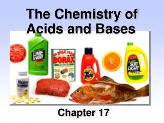
Buffering Systems • A buffer is a chemical that can bind excessive H+ or OH– without a significant change in pH • A buffering pair consists of a weak acid and its conjugate base • The most important plasma buffering systems are the carbonic acid–bicarbonate system and hemoglobin
Carbonic Acid–Bicarbonate Pair • Operates in both the lung and the kidney • The greater the partial pressure of carbon dioxide, the more carbonic acid is formed • At a pH of 7.4, the ratio of bicarbonate to carbonic acid is 20:1 • Bicarbonate and carbonic acid can increase or decrease, but the ratio must be maintained
Carbonic Acid–Bicarbonate Pair • If the amount of bicarbonate decreases, the pH decreases, causing a state of acidosis • The pH can be returned to normal if the amount of carbonic acid also decreases • This type of pH adjustment is referred to as compensation • The respiratory system compensates by increasing or decreasing ventilation • The renal system compensates by producing acidic or alkaline urine
Other Buffering Systems • Protein buffering • Proteins have negative charges, so they can serve as buffers for H+ • Renal buffering • Secretion of H+ in the urine and reabsorption of HCO3– • Cellular ion exchange • Exchange of K+ for H+ in acidosis and alkalosis
Acid-Base Imbalances • Normal arterial blood pH • 7.35 to 7.45 • Obtained by arterial blood gas (ABG) sampling • Acidosis • Systemic increase in H+ concentration • Alkalosis • Systemic decrease in H+ concentration
Acidosis and Alkalosis • Four categories of acid-base imbalances: • Respiratory acidosis—elevation of pCO2 due to ventilation depression • Respiratory alkalosis—depression of pCO2 due to alveolar hyperventilation • Metabolic acidosis—depression of HCO3– or an increase in non-carbonic acids • Metabolic alkalosis—elevation of HCO3– usually due to an excessive loss of metabolic acids
Anion Gap • Used cautiously to distinguish different types of metabolic acidosis • By rule, the concentration of anions (–) should equal the concentration of cations (+). Not all normal anions are routinely measured. • Normal anion gap = Na+ + K+= Cl– + HCO3– + 10 to 12 mEq/L (other misc. anions [the ones we don’t measure]—phosphates, sulfates, organic acids, etc.)
Anion Gap • An abnormal anion gap occurs due to an increased level of an abnormal unmeasured anion • Examples: DKA—ketones, salicylate poisoning, lactic acidosis—increased lactic acid, renal failure, etc. • As these abnormal anions accumulate, the measured anions have to decrease to maintain electroneutrality