Download
1 / 70
760 likes | 1.51k Views
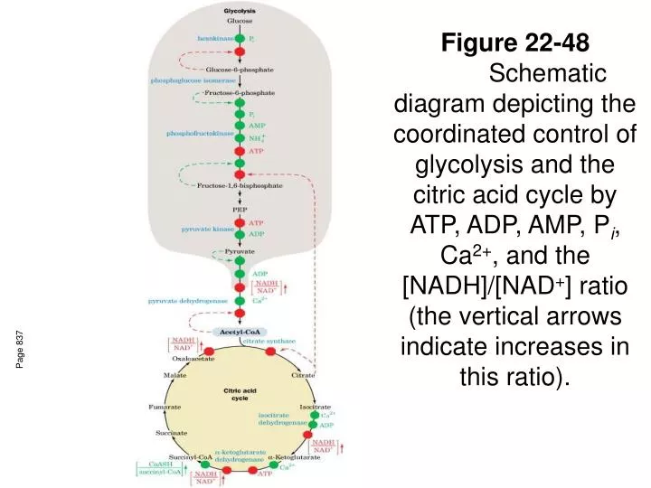
Figure 22-48 Schematic diagram depicting the coordinated control of glycolysis and the citric acid cycle by ATP, ADP, AMP, P i , Ca 2+ , and the [NADH]/[NAD + ] ratio (the vertical arrows indicate increases in this ratio). Page 837. CH 23: Gluconeogenisis and Pentose Phosphate Pathway.

E N D
Figure 22-48 Schematic diagram depicting the coordinated control of glycolysis and the citric acid cycle by ATP, ADP, AMP, Pi, Ca2+, and the [NADH]/[NAD+] ratio (the vertical arrows indicate increases in this ratio). Page 837
CH 23: Gluconeogenisis and Pentose Phosphate Pathway Much as I hate to skip stuff in this chapter, we will cover pp 843-850 and 862-870. Please read section 3 on glycoprotein synthesis, pp 852-861. You should be able to do all the problems…
Gluconeogenesis • This route is important when fasting • Precursors: lactate, pryuvate, TCA intermediates, most aa’s (except leu,lys) • Entry into gluconeogenisis: OAA • Note that animals cannot make glucose from AcetylCoA (plants have the glyoxylate cycle)
Figure 23-1 Pathways converting lactate, pyruvate, and citric acid cycle intermediates to oxaloacetate. Page 844
Synthesis and degradation are always separated • The really good news: Mostly glycolytic enzymes involved. • What irreversible enzymes of glycolysis must be bypassed for gluconeogenesis???? • PK, PFK, HK
Figure 23-2 Conversion of pyruvate to oxaloacetate and then to phosphoenolpyruvate. Prosthetic group=biotin Page 845 Hi energy intermediate
Figure 23-3aBiotin and carboxybiotinyl–enzyme. (a) Biotin consists of an imidazoline ring that is cis-fused to a tetrahydrothiophene ring bearing a valerate side chain. Raw eggs contain avidin—a protein with very high affinity for biotin Bacteria (Streptomyces) make avidin analogs like streptavidin—where did we see this recently??? http://www.rpi.edu/dept/bcbp/molbiochem/MBWeb/mb1/part2/chime/biotin/btn-index.html
Figure 23-3b Biotin and carboxybiotinyl–enzyme. (b) In carboxybiotinyl–enzyme, N1 of the biotin ureidogroup is the carboxylation site. Another swinging arm between 2 acitive sites of enzyme! Page 845
Figure 23-4 Two-phase reaction mechanism of pyruvate carboxylase. Phase 1: carboxylation of biotin
Figure 23-4 (continued) Two-phase reaction mechanism of pyruvate carboxylase. Phase II: carboxylation of pyruvate PEP nucleophillically attacks CO2 Page 846 Biotin accepts H+ from pyruvate→PEP CO2 produced in active site via elimination
Pyruvate Carboxylase Facts • Catalyzes an important anaplerotic reaction that increases TCA activity • Acetyl-CoA allosterically activates PC* • *If TCA is inhibited by hi ATP/NADH, OAA →gluconeogenesis
Figure 23-5 The PEPCK mechanism. GTP driven decarboxylation of OAA →PEP
Figure 23-6 Transport of PEP and OAA from the mitochondria to the cytosol. 2 different routes—either via malate or asp Malate shuttle also moves NADH (required in cytosol for gluconeo-genesis Page 847
Glc-6-Phosphatase unique to kidney and liver They supply other tissues with glc. Figure 23-7 Pathways of gluconeogenesis and glycolysis. Page 848
Table 23-1 Regulators of Gluconeogenic Enzyme Activity. Page 849
Figure 23-9 The Cori cycle. Page 850
Cells’ second energy currency: NADPH! • NADPH is required for reductive biosynthesis • FA’s • Steroids • Photosynthesis • Etc. • NADPH is generated by oxidation of G6P • Pentose phosphate pathway (PPP) = hexose monophosphate shunt= phosphogluconate pathway • 3 G6P + 6 NADP+ + 3 H2O →6 NADPH + 6 H+ + 3 CO2 + 2 F6P = GA3P • Pathway divided into 3 phases • Oxidative Reactions • Produces Ribulose-5-P • Isomerization/Epimerization Reactions • Produces Ribose-5-P and Xyulose-5-P • Transaldolase and Transketolase Reactions • 3 Ru5P ↔r5P + 2 Xu5P Most cells maintain their [NAD+]/[NADPH] near 1000!!!
Figure 23-26 The glucose-6-phosphate dehydrogenase reaction. Page 864
Figure 23-27 The phosphogluconate dehydrogenase reaction. Page 864
Figure 23-28 Ribulose- 5-phosphate isomerase and ribulose- 5-phosphate epimerase. Page 865
Figure 23-31 Summary of carbon skeleton rearrangements in the pentose phosphate pathway. Page 867
CH 23: Gluconeogenisis and Pentose Phosphate Pathway
Figure 23-1 Pathways converting lactate, pyruvate, and citric acid cycle intermediates to oxaloacetate. Page 844
Figure 23-2 Conversion of pyruvate to oxaloacetate and then to phosphoenolpyruvate. Page 845
Figure 23-3a Biotin and carboxybiotinyl–enzyme. (a) Biotin consists of an imidazoline ring that is cis-fused to a tetrahydrothiophene ring bearing a valerate side chain.
Figure 23-3b Biotin and carboxybiotinyl–enzyme. (b) In carboxybiotinyl–enzyme, N1 of the biotin ureido group is the carboxylation site. Page 845
Figure 23-4 Two-phase reaction mechanism of pyruvate carboxylase. Page 846
Figure 23-4 (continued) Two-phase reaction mechanism of pyruvate carboxylase. Phase II Page 846
Page 847 Figure 23-5 The PEPCK mechanism.
Figure 23-6 Transport of PEP and OAA from the mitochondrion to the cytosol. Page 847
Figure 23-7 Pathways of gluconeogenesis and glycolysis. Page 848
Table 23-1 Regulators of Gluconeogenic Enzyme Activity. Page 849
Figure 23-26 The glucose-6-phosphate dehydrogenase reaction. Page 864
Figure 23-27 The phosphogluconate dehydrogenase reaction. Page 864
Figure 23-28 Ribulose- 5-phosphate isomerase and ribulose- 5-phosphate epimerase. Page 865
Figure 23-31 Summary of carbon skeleton rearrangements in the pentose phosphate pathway. Page 867
Figure 24-1 Chloroplast from corn. Page 872 Photosynthesis!!! Ch 24
Figure 24-3 Chlorophyll structures. Page 874
Figure 24-4 Energy diagram indicating the electronic states of chlorophyll and their most important modes of inter-conversion. Page 875
Figure 24-5 Absorption spectra of various photosynthetic pigments. Page 875
Figure 24-7a Flow of energy through a photosynthetic antenna complex. (a) The excitation resulting from photon absorption randomlymigrates by exciton transfer. Page 877