Download

1 / 47
600 likes | 1.3k Views
Methods for Structure Determination. Chemistry and Chemical Biology Rutgers University. How are macromolecular structures determined?. X-ray (X-ray crystallography). NMR (Nuclear Magnetic Resonance). EM (Electron Microscopy). Protein Data Bank. Download. X-ray Crystallography.

E N D
Methods for Structure Determination Chemistry and Chemical Biology Rutgers University
How are macromolecular structures determined? X-ray (X-ray crystallography) NMR (Nuclear Magnetic Resonance) EM (Electron Microscopy) Protein Data Bank Download
X-ray Crystallography Target selection Protein production Crystallization Structure calculation Refinement Validation Deposition to PDB Annotation Data collection Phasing http://www-structmed.cimr.cam.ac.uk/Course/Overview/Overview.html#methods
Some Common Concepts • Symmetry: Translation, Rotation, Reflection, Inversion • Crystals: Lattice, Unit cell, Asymmetric Unit • Diffraction: Light diffraction, X-ray diffraction, diffraction patterns • Real vs Reciprocal space: Fourier transform • Phase problem: What is it? Phasing strategies • Structure solution pipeline
Symmetry Translation Rotation (pt) Inversion?? (pt) Mirror (pt) M.C. Escher
Mineral Protein Crystals
, , , , Unit Cell 1 , , , , , , , , , , , , , , , , , , , , , , , , Convolution , , , , , , , , , , , , Crystal structure , , , , , , , , Unit Cell 2 Lattice, Crystal and Unit cell lattice . . . . . . . . . . . . . . . . , object
Macromolecular Crystal Lattice Alexander McPherson, Introduction to Macromolecular Crystallography Wiley-Liss, 2002
Symmetry in Crystals 1 • 1-fold • 2-fold • 3-fold • 4-fold • 6-fold 2 3 4 5-, 7-, 8- and higher fold symmetries 6 do not pack in a crystal
Crystal Systems Z. Dautera & M. Jaskolskib http://journals.iucr.org/j/issues/2010/05/02/kk5061/kk5061bdy.html
Diffraction Sunrise through a screened window http://www.flickr.com/photos/fizzix/2458009067/in/photostream/
Diffraction in Action http://mrsec.wisc.edu/Edetc/supplies/DNA_OTK/images/ABCH.mov
Vs Direct/Real space Reciprocal space Diffraction pattern Reciprocal lattice (a*, b*, c*, diffraction spot distance d*) Structure factor • Crystal • Crystal lattice (a,b,c, planes distance d) • Electron density Fourier Transform http://www.nist.gov/lispix/doc/menu-bar/fft-tool.htm Try out an interactive tutorial at: http://www.lks.physik.uni-erlangen.de/diffraction/space.html
The Fourier Duck Fourier Transform Reverse Transform Reverse Transform with limited resolution data
Why Use X-rays? http://bccp.lbl.gov/Academy/wksp_pix_1/spectrum.gif
X-ray Diffraction Gale Rhodes, Crystallography Made Crystal Clear: A Guide for Users of Macromolecular Models, Academic Press, 1993
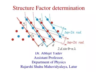
Bragg’s Law • n = 2d sin • 2 angle between incident • and reflected beams d spacing between planes • wavelength n order of diffraction http://www.bmsc.washington.edu/people/merritt/bc530/bragg/ try the Java Applet! Constructive interference occurs from successive crystallographic planes (h, k, l) in the crystalline lattice
Microscopy vs X-ray Crystallography http://www.iucr.org/education/pamphlets/15/full-text
The Phase Problem http://www.ruppweb.org/Xray/Phasing/Phasingt.html
The X-ray Crystallography Pipeline Crystal growth Data collection Phase determination Model building and refinement Protein preparation
Crystal Growth: Vapor Diffusion Cover Slip Precipitant Solution Protein + Precipitant Common precipitants: • Polyethylene glycol • Salts • ammonium sulfate • sodium chloride • Alcohols • Isopropanol • Methylpentanediol (MPD)
Data Collection Crystal mounted in glass capillary Crystal mounted in nylon loop. Frozen in liquid N2 Rotating Anode Diffractometer
Synchrotron X-ray source http://www.nsls.bnl.gov NSLS Beamline X12C
Crystal Diffraction High Resolution (large angle) Water Ring ~3-5 Å Beam Stop Shadow Low Resolution (small angle) Jeff Dahl, Sars protease, http://en.wikipedia.org/wiki/File:X-ray_diffraction_pattern_3clpro.jpg
trp repressor, sodium phosphate trp repressor, ammonium sulfate Different crystal forms of the same protein yield different diffraction patterns
X-ray Diffraction Pattern • Diffraction pattern is in reciprocal space • Size and shape of unit cell determines position of diffraction peaks. • Atomic positions within unit cell determines intensity of peaks. • Experimental data: h,k,l and intensities (with errors) A precession photograph
Data Obtained a = 36.67 Å b = 79.39 Å c = 39.97 Å α = 90.0° ß = 91.25° γ = 90.0° Monoclinic lattice (P2 or P21) H K L intensity error 0 0 12 6714.3 347.2 0 0 18 -8.9 16.3 0 0 24 979.5 62.4 0 0 30 4136.4 272.5 1 0 3 3035.4 70.2 1 0 4 0.0 0.7 1 0 5 0.1 0.6 1 0 6 838.4 20.4 1 0 7 14903.0 535.6 1 0 8 2759.4 64.7 1 0 9 1403.5 31.0 1 0 10 109.4 5.6 1 0 11 31739.5 1611.5 1 0 12 231.9 7.6 ...etc. Crystal unit cell dimensions Lattice type, possible space groups Resolution Limit Merged data set with index, intensity + error for each reflection
Ihkl=constant.|Fhkl|2 Structure Factor Structure Factor r(x,y,z) =ΣFhkle-2πi (hx + ky +lz) Electron Density Diffraction Patterns to Structure
Phase Problem • Structure factor is dependent on type and location of atoms in unit cell • The complete Structure Factor Ffor a reflection includes the phase, which cannot be measured directly. Fhkl = |Fhkl|eiϕhkl Structure Factor Phase: must be estimated Amplitude: from experimental measurements
Phase Determination • Direct methods • Estimate from probability relationships applied to most intense diffraction peaks • Patterson methods • Multiple Isomorphous Replacement • Anomalous Dispersion • Molecular replacement • Density Improvement • Non-crystallographic symmetry averaging • Solvent flattening
Patterson Function • Convolution of electron density with itself • Evaluated at points u,v,w throughout unit cell • Map of vectors between scattering atom in the real crystal cell (translated to Patterson origin) Patterson map crystal http://www.ruppweb.org/Xray/Patterson/Native_Patterson.htm
Isomorphous Replacement • Derivative – native crystal = heavy atom • Deriv. diffn – native diffn = heavy atom diffn • Patterson synthesis > peaks based on distance between heavy atoms in structure gives initial phase. Real space Reciprocal space http://www.ruppweb.org/Xray/Phasing/Phasingt.html
Anomalous Dispersion • Friedel’s Law: Ihkl = I-h-k-l • Members of a Friedel pair have equal amplitude and opposite phase • In anomalous scattering crystals Friedel’s law is not obeyed http://www.xtal.iqfr.csic.es/Cristalografia/parte_07_2-en.html http://skuld.bmsc.washington.edu/scatter/AS_wavechoice.html
Molecular Replacement • New structure expected to resemble one previously determined • Use Patterson-based methods to find the orientation of known model in new crystal lattice (i.e. find rotation R and translation T) http://reference.iucr.org/dictionary/Molecular_replacement
Electron Density • Can be calculated by Fourier transform of diffraction data • Provides an averaged image: • over all molecules in the crystal • over the time of the diffraction experiment Trp in a 4.3 A map Trp in a 1.3 A map Trp in a 2.25 A map
Model Building-Refinement Cycle Final Model
-snip- Structural Data PDB 3a6b
Types of Electron Density Maps • Experimentally phased map: • Fobs, Phicalc • “model” map: • (2Fobs – Fcalc), Phicalc • “difference” map • (Fobs – Fcalc) or (Fobs – Fobs), Phicalc
Summary: The Data Pipeline Isolation, Expression, Purification, Crystallization Genomic Based Target Selection Data Collection Structure Determination PDB Deposition & Release X-ray cryst NMR 3D Models Annotations Publications EM
Some Movie Links • Crystal Mounting Robot • http://www.youtube.com/watch?v=J4OD_b9XKh4 • Crystal Diffraction • http://ucxray.berkeley.edu/~jamesh/movies/ • Optical diffraction • http://mrsec.wisc.edu/Edetc/supplies/DNA_OTK/index.html Enjoy!
References • IUCr Online dictionary of Crystallography • http://reference.iucr.org/dictionary/Main_Page • Educational web sites and resources • http://www.iucr.org/education/resources • An interactive SF tutorial • http://www.ysbl.york.ac.uk/~cowtan/sfapplet/sfintro.html