Download
1 / 1
10 likes | 204 Views
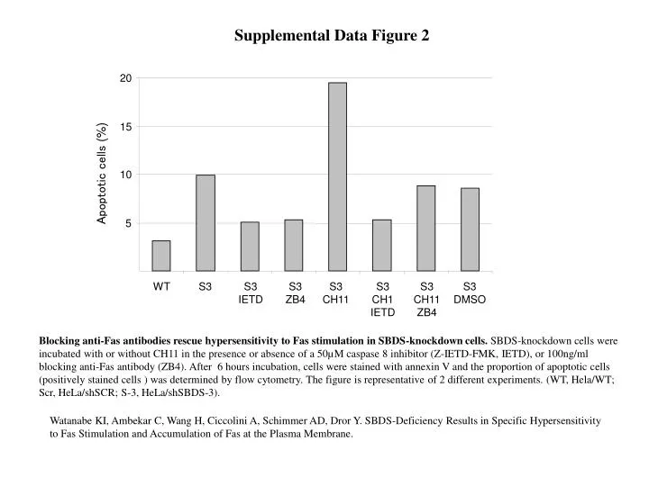
Supplemental Data Figure 2. 20. 15. Apoptotic cells (%). 10. 5. WT. S3. S3 IETD. S3 ZB4. S3 CH11. S3 CH1 IETD. S3 CH11 ZB4. S3 DMSO.

E N D
Supplemental Data Figure 2 20 15 Apoptotic cells (%) 10 5 WT S3 S3 IETD S3 ZB4 S3 CH11 S3 CH1 IETD S3 CH11 ZB4 S3 DMSO Blocking anti-Fas antibodies rescue hypersensitivity to Fas stimulation in SBDS-knockdown cells. SBDS-knockdown cells were incubated with or without CH11 in the presence or absence of a 50µM caspase 8 inhibitor (Z-IETD-FMK, IETD), or 100ng/ml blocking anti-Fas antibody (ZB4). After 6 hours incubation, cells were stained with annexin V and the proportion of apoptotic cells (positively stained cells ) was determined by flow cytometry. The figure is representative of 2 different experiments. (WT, Hela/WT; Scr, HeLa/shSCR; S-3, HeLa/shSBDS-3). Watanabe KI, Ambekar C, Wang H, Ciccolini A, Schimmer AD, Dror Y. SBDS-Deficiency Results in Specific Hypersensitivity to Fas Stimulation and Accumulation of Fas at the Plasma Membrane.