Download
1 / 4
71 likes | 491 Views
Lymphomatoid papulosis – a case report Dr Georgescu Mihaela*, Dr Margaritescu Irina** *SUUMC “Carol Davila”, Bucharest, ** Domina Sana Medical Center, Bucharest. Clinical case A 18 years old female presented with a 3 years-history of polymorphic skin lesions, at inferior limbs

E N D
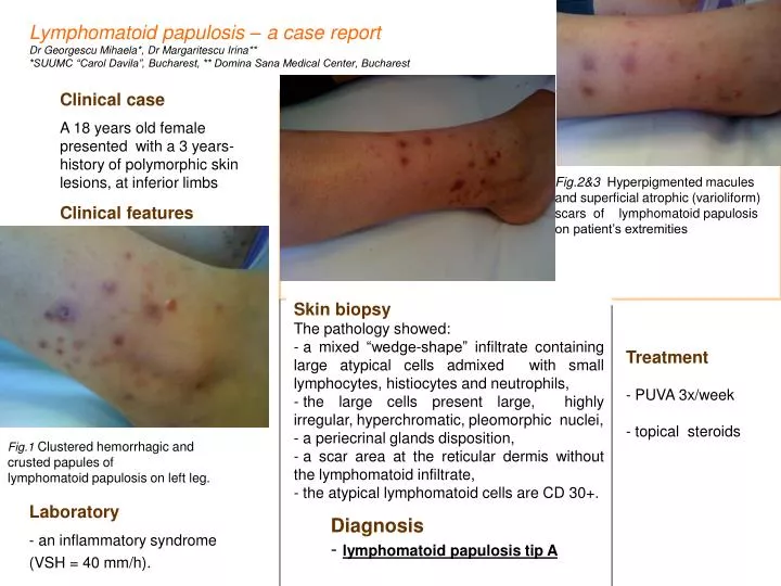
Lymphomatoid papulosis – a case report Dr Georgescu Mihaela*, Dr Margaritescu Irina** *SUUMC “Carol Davila”, Bucharest, ** Domina Sana Medical Center, Bucharest Clinical case A 18 years old female presented with a 3 years-history of polymorphic skin lesions, at inferior limbs Clinical features Fig.2&3Hyperpigmented macules and superficial atrophic (varioliform)scars of lymphomatoid papulosis on patient’s extremities • Skin biopsy • The pathology showed: • a mixed “wedge-shape” infiltrate containing large atypical cells admixed with small lymphocytes, histiocytes and neutrophils, • the large cells present large, highly irregular, hyperchromatic, pleomorphic nuclei, • a periecrinal glands disposition, • a scar area at the reticular dermis without the lymphomatoid infiltrate, • the atypical lymphomatoid cells are CD 30+. Treatment - PUVA 3x/week - topical steroids Fig.1Clustered hemorrhagic and crusted papules of lymphomatoid papulosis on left leg. Laboratory - an inflammatory syndrome (VSH = 40 mm/h). Diagnosis - lymphomatoid papulosis tip A
Histopathology Lymphomatoid papulosis – a case report Dr Georgescu Mihaela*, Dr Margaritescu Irina** *SUUMC “Carol Davila”, Bucharest, ** Domina Sana Medical Center, Bucharest Fig.4 HEx50 Figure 5. HEx200 Figure 6. HEx400 Figure 4,5 Fragment of cutaneous biopsy withmixed “wedge-shape” infiltrate containing large atypical cells admixed with small lymphocytes, histiocytes and neutrophils Figure 6. Lymphomatoid papulosis type A lesion with predominance of large atypicallymphoid cells, displaying polymorphic nuclei, prominent nucleoli, and abundant cytoplasm Figure 7,8 Numerous CD 30+ cells are present. Figure 7. HEx200 Figure 8. HEx400
Lymphomatoid papulosis – a case report Dr Georgescu Mihaela*, Dr Margaritescu Irina** *SUUMC “Carol Davila”, Bucharest, ** Domina Sana Medical Center, Bucharest Introduction Lymphomatoid papulosis is a peculiar condition, caractherized by a chronic, recurrent, self-healing papulonecrotic or papulonodular eruption with histopathological features suggestive of a (CD 30 – positive) malignant lymphoma The pathogenesis • unknown • a possible viral-driven etiology • a low-grade lymphoma induced by chronic antigenic stimulation • studies show a high rate of apoptosis contributing to regression, mediated by death-receptor pathway signaling via cell surface Fas (CD95) signaling and/or due to increased levels of the proapoptotic protein bax. Mutations of TGF-β signaling receptor genes results in disease progression Differential diagnosis • viral infections (herpes virus, molluscum contagiosum, parapox virus (milker's nodule), Epstein-Barr virus, HTLV1, HIV ) • scabies • syphilis • superficial fungal infections • pityriais lichenoides et varioliformis acuta • atopic dermatitis • drug reactions (particularly to anticonvulsants) • mycosis fungoides or a lymphomatoid drug reaction (type B lymphomatoid papulosis) • Treatment • - active treatment -not necessary(few non-scarring lesions) • - no treatment has proven consistently effective • - beneficial effects from: • PUVA • topical mechlorethamine or carmustine • low-dose etoposide • Clinical features • typicalskin lesions are red-brown papules and nodules that may develop central hemorrhage, necrosis and crusting, which disappear within 3-8 weeks • characteristically, lesions are often at different stages • of development
Lymphomatoid papulosis – a case report Dr Georgescu Mihaela*, Dr Margaritescu Irina** *SUUMC “Carol Davila”, Bucharest, ** Domina Sana Medical Center, Bucharest Histological types Differential diagnosis Type A - Scattered CD30+, large Hodgkin lymphoma Type B - Epidermotropic CD30-/+ small Mycosis fungoides Type C - Cohesive sheets CD30+, large ALCL Type D - Epidermotropic CD30+ CD8+ small AECTCL (Berti lymphoma) Type E - Angioinvasive CD30+ CD8+>CD4+Extranodal NK/T, GD-TCL References 1. McKee’s Pathology of the Skin, Chapter 29 – Cutaneous lymphoproliferative diseases and related disorders, John Goodlad,Eduardo Calonje, Lymphomatoid papulosis, 4th edition, Elsevier, 2012 2. Skin Lymphoma: The Illustrated Guide, Lorenzo Cerroni et al, CD30+ lymphoproliferative disorders. Lymphomatoid papulosis3rd Edition, Wiley, 2009 3. Dermatology, By By Jean L. Bolognia et al., Chapter 120. Primary cutaneous CD30-positive lymphoproliferative disorder. Lymphomatoid papulosis, 3rd Edition, Elsevier, 2012 4. Self assessmentCourse in Virtual Dermatopathology, Dr. Werner Kempf, Case 02, EADV Congress, Istanbul, 2-6 oct 2013 Conclusions • The disease is now classified as an indolent lymphoma in the new WHO classification • The clinicopathological and immunohistochemical correlation is essential in establishing the diagnosis The evolution of the disease is characterized by recurrence