Download
1 / 37
380 likes | 957 Views
Alveolar Process. Gingiva. Near the end of the 2nd month of fetal life, mandible and maxilla form a groove that is opened toward the surface of the oral cavity As tooth germs start to develop, bony septa form gradually. The alveolar process starts developing strictly during tooth eruption.

E N D
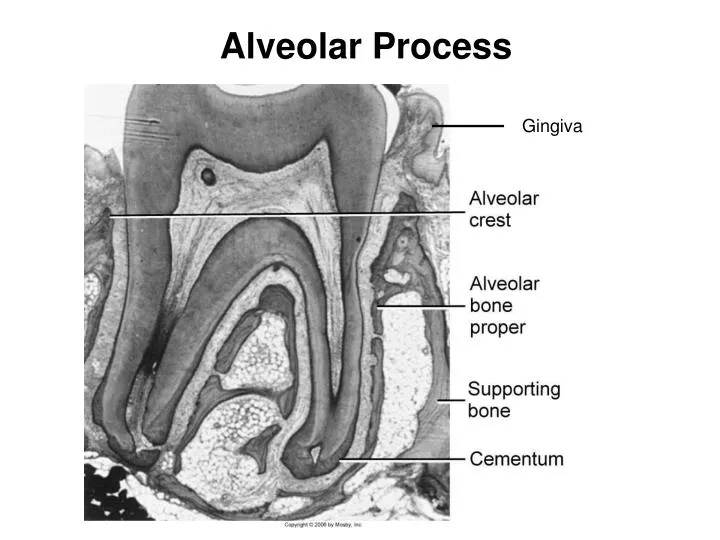
Alveolar Process Gingiva
Near the end of the 2nd month of fetal life, mandible and maxilla form a groove that is opened toward the surface of the oral cavity As tooth germs start to develop, bony septa form gradually. The alveolar process starts developing strictly during tooth eruption.
a) outer cortical platesb) a central spongiosa c) bone lining the alveolus (bundle bone)
Alveolar bone proper: The compact or dense bone that lines the tooth. Contains either perforating fibers from periodontal ligament (Sharpey’s fibers) or just compact bone Sharpey’s fibers embedded into the alveolar bone proper Present at right angles or oblique to the surface of alveolar bone and along the root surface Because alveolar process is regularly penetrated by collagen fiber bundles, it is also called bundle bone. It appears more radiodense than surrounding supporting bone in X-rays called lamina dura
Bundle Bone It is perforated by many foramina that transmit nerves and vessels (cribriform plate). Radiographically, the bundle bone is the lamina dura. The lining of the alveolus is fairly smooth in the young but rougher in the adults. Radiodense because increased mineral content around fiber bundles Lamina Dura
Supporting Compact Bone Similar to compact bone anywhere else (Haversian bone) Extends both on the lingual (palatal) and buccal side Contains haversian and Volkman’s canals (they both form a continuous channel of nutrient canals)
Bundle bone and Trabecular bone Arrows: Sharpey’s fiber
The alveolar crest is found 1.5-2.0 mm below the level of the CEJ. If you draw a line connecting the CE junctions of adjacent teeth, this line should be parallel to the alveolar crest. If the line is not parallel, then there is high probability of periodontal disease.
Clinical considerations Resorption and regeneration of alveolar bone This process can occur during orthodontic movement of teeth. Bone is resorbed on the side of pressure and opposed on the site of tension. Decreased bone (osteopenia) of alveolar process is noted when there is inactivity of tooth that does not have an antagonist
Periodontal Ligament PDL is the soft specialized connective tissue situated between cementum and alveolar bone proper Ranges in thickness between 0.15 and 0.38 mm and is thinnest in the middle portion of the root The width decreases with age Tissue with high turnover rate Contains fibers, cells and intercellular substance
Embryogenesis The PDL forms from the dental follicle shortly after root development begins
FUNCTIONS OF PERIODONTIUM Tooth support Shock absorber: Withstanding the forces of mastication Sensory receptor necessary for proper positioning of the jaw Nutritive: blood vessels provide the essential nutrients to the vitality of the PDL
Cells a) Osteoblasts b) Osteoclasts (critical for periodontal disease and tooth movement) c) Fibroblasts (Most abundant) d) Epithelial cells (remnants of Hertwig’s epithelial root sheath-epithelial cell rests of Malassez) e) Macrophages (important defense cells) f) Undifferentiated cells (perivascular location)h) Cementoblasts i) Cementoclasts (only in pathologic conditions)
PDL fibers • Collagen fibers: I, III and XII. Groups of fibers that are continually remodeled. (Principal fiber bundles of the PDL). The average diameter of individual fibers are smaller than other areas of the body, due to the shorter half-life of PDL fibers (so they have less time for fibrillar assembly) • - Oxytalan fibers: variant of elastic fibers, perpendicular to teeth, adjacent to capillaries • - Eluanin: variant of elastic fibers
Principal Fibers Run between tooth and bone. Can be classified as dentoalveolar and gingival group Dentoalveolar group a. Alveolar crest group (ACG): below CE junction, downward, outwardb. Horizontal group: apical to ACG, right angle to the root surface c. Oblique group: most numerous, oblique direction and attaches coronally to boned. Apical group: around the apex, base of sockete. Interradicular group: multirooted teeth Runs from cementum and bone , forming the crest of the interradicular septum At each end, fibers embedded in bone and cementum: Sharpey’s fiber
Gingival ligament fibers: the principal fibers in the gingival area are referred to as gingival fibers. Not strictly related to periodontium. Present in the lamina propria of the gingiva. a. Dentogingival: most numerous; cervical cementum to f/a gingivab. Alveologingival: bone of the alveolar crest to f/a gingivac. Circular: around neck of teeth, free gingivad. Dentoperiosteal: runs apically from the cementum over the outer cortical plate to alv. process or vestibule (muscle) or floor of mouthe. Transseptal: cementum between adjacent teeth, over the alveolar crest
Transeptal Alveolar crest Horizontal Oblique
Oxytalan Fibers Type of elastic fibers present as bundes of microfibrils that run oblique from the cementum surface to the blood vessels. Associated with neural elements. Most numerous in the cervical area. Function: Regulate vascular flow in relation to tooth function
The PDL gets its blood supply from perforating arteries (from the cribriform plate of the bundle bone). The small capillaries derive from the superior & inferior alveolar arteries. The blood supply is rich because the PDL has a very high turnover as a tissue. The posterior supply is more prominent than the anterior. The mandibular is more prominent than the maxillary.
Nerve supply The nerve supply originates from the inferior or the superior alveolar nerves. The fibers enter from the apical region and lateral socket walls. The apical region contains more nerve endings (except Upper Incisors)
Interstitial Space Present between each bundle of ligament fibers Contains blood vessels and nerves Designed to withstand the impact of masticatory forces
Ground Substance Amorphous background material that binds tissues and fluids A major constituent of the PDL Similar to most connective tissue ground substance Dermatan sulfate is the major glycosaminoglycan 70% water; critical for withstanding forces When function is increased PDL is increased in size and fiber thickens Bone trabeculae also increase in number and thicker However, in reduction of function, PDL narrows and fiber bundles decreases in number and thickness (this reduction in PDL is primarily due to increased cementum deposition)