Download
1 / 56
720 likes | 1.58k Views
Phylum: Platyhelminthes (Flatworms) 1-Cestoda (tape worms). 2-Trematoda (flukes). Cestoda (tape worms) A) Intestinal adult tapeworms. B) Extra-intestinal larval tapeworms. Class: Cestoda (Tape worm) 1- The length of the different species varies from 3mm to 10 meters.

E N D
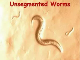
Phylum: Platyhelminthes (Flatworms) 1-Cestoda (tape worms). 2-Trematoda (flukes). Cestoda (tape worms) A) Intestinal adult tapeworms. B) Extra-intestinal larval tapeworms.
Class: Cestoda (Tape worm) 1- The length of the different species varies from 3mm to 10 meters. 2-Segmented body: The body of the typical cestode consists of 3 distinct regions: scolex, neck, and strobila 3- Scolex (head) provided with suckers, sometime hooks. 4- Hermaphroditic mature segment has male and female reproductive system 5- Despite the lack of a digestive system (no mouth, no gut, no anus) they do absorb food from the hosts intestine
General Morphology • The body of the typical cestode consists of 3 distinct regions: • scolex • neck • strobila
Form and Function: The Scolex cont. • The scolices of tapeworms are typically categorized as either acetabulate or bothriate, depending on the type of sucker present • An acetabulate scolex is characterized by the presence of 4 muscular cups sunk into the equatorial surface of the scolex; cups are radially arranged equidistant from each other • In addition to muscular cups, there may be accessory holdfast structures, such as hooks to help anchor the scolex to the host’s intestinal wall • In this case, the scolex is called an armed scolex • These hooks are usually grouped at the apical end of the scolex on a protrusible rostellum rostellum
Scolex: is located at the anterior end and functions as an attachment structure • 4 suckers • hooklets:These hooks are usually grouped at the apical end of the scolex on a protrusiblerostellum
Neck • The neck is an unsegmented, poorly differentiated region immediately posterior to the scolex • short measuring 5 to 10 mm in length
The Strobila • As new proglottids are formed from the neck region, they push the older ones progressively posteriad, creating a chain of proglottids - the strobila • The asexual process of forming segments is termed strobilation. The stroblia can be loosely subdivided into 3 regions: • Immature • Mature • Gravid proglottids
mature segment contain testes and ovary
gravid proglottid has a median uterus filled with eggs (50 – 80 000)
Larval (metacestoda): 1- The larval are extracellular parasites, visible to the naked eye, seen as bladders with an scolex. 2- The external surface is a tegumentary tissue, similar to that found in the adult worm. 3- In human infection, these larvae can survive for a many years. 4- Dead parasite tissue, leaving a calcified in both muscle and brain tissue.
Intestinal adult tapeworms. Taenia solium Taenia saginata Taenia multicepsisHymenolepis nanaHymenolepis diminuta Dipylidium caninumDiphyllobothrium latum Extra-intestinal larval tapeworms Echinococcus granulosus Echinococcus multilocularis
Taenia solium • Common name: The Pork Tapeworm. • Habitat: Small intestine. • Route of infection: Eating of unwell cooked Pork meat. • Definitive host : Human. • Intermediate host: Pork, Human (occasional). • Infective stage: Cysticercus larvae (Pork). • Diagnostic stage: Eggs or gravid in Feces. • Disease:Adults cause: Taeniasis. Larvae cause: Cysticercosis( presences of Cysticercus larvae in brain and muscles.
Taenia solium Adult worm 1- 2-4 meters in length) 2- Head or scolex globular in shaped with 4 suckers rounded rostellum armed with double rows of large and small hooks numbering 22 to 36. 3- Neck short measuring 5 to 10 mm in length.
Proglottids • Numbers : 700 to 1000 proglottids Composed of: 1- Immature proglottid 2- Mature proglottid: nearly square containing full set of functioning male and female reproductive organs 3- Gravid proglottid • longer than broader consists: • gravid uterus with 3 to 13 lateral uterine branches arranged.
Egg • Shape – spherical • Size: 30-40 um in diameter • radially-striated shells • Outer shell – thin and rarely seen • Inner shell brown, thick and striated • embryo or oncosphere with six hooklets `
Larval stage or bladder worm • also called Cysticercus cellulosae • measurement – 5 to 10 mm in length and 5 mm in diameter • scolex • hooks • suckers.
EPIDEMIOLOGY T. solium infection • Human infected (raw or undercooked pork) • Man is the only definitive host and the pig appears to be the only intermediate host Man become the intermediate host Can be caused by: • ingestion of eggs from contaminated food or water • by internal autoinfection when the eggs are carried by reverse peristalsis back to the duodenum or stomach
Pathology 1- By adult in lumen of the small intestines : • intestinal obstruction • abdominal pain • Vomiting . • nausea • weight loss and diarrhea.
2- By larval stage (cysticercus) Symptoms depend on location and number of larva, which encyst in the muscle and other tissues : a. cellular reactions b. fibrosis c. necrosis • cysticercosis in the brain may cause: a. epilepsy b. meningitis, and encephalitis • Eye – cause blindness
Diagnosis: 1-Adult worms: Taenia infections are diagnosis by finding gravid segments in the feces, because their eggs are identical. 2- Cysticercosis: A- Serologic tests ELISA. B- X-rays may reveal calcified cysticerci. C- CT scan can show living cysticerci.
Taenia saginata Beef Tapeworm.
Taenia saginata • Common name: The Beef Tapeworm. • Habitat: Small intestine. • infection: Eating of unwell cooked cow meat. • Definitive host : Human. • Intermediate host: Cows also other herbivores. • Infective stage: Cysticercus larvae. • Diagnostic stage: Eggs and gravid in Feces. • Disease:Adults cause Taeniasis.
Taenia soliumTaenia saginata Intermediate host: Pig cattle Length : 2-4 m 4-8 m No. proglottids: 700-1000 1000-2000 Scolex : 4 suckers 4 suckers but with hooklets no hooklets
Taenia soliumTaenia saginata Gravid proglottid: 3-13 branches 13-30 branches
Larval stage (Cysticercus) Taenia soliumTaenia saginata (Cysticercus cellulose)(Cysticercus bovis) Scolex with hooklets no hooklets on Scolex found in pig and man only found in cattle
Echinococcus granulosus Dog tape worm
Echinococcus granulosus Dog tape worm • Habitat: adult: Small intestine in Dogs Larvae (Hydatid cyst): in Liver, Lung, brain in Intermediate host • Definitive host : Dogs and other canines. • Intermediate host: Sheep, cattle, etc, and other herbivores. Accidentally Human. • Disease: Unilocular Hydatid disease.
Morphology: • 3-8 mm in length. • Scolex with Four suckers. • Neck. • Immature segment. • Mature segment. • Gravid segment.
Ova: • Shape Round to Oval • Embryonated ( Hexacanth embryo inside) • Radially striated egg shell. • Onchosphere: six-hooked embryo inside the egg.
Hydatid Cyst: • At maturity, the cyst wall contains 2 layers: • 1- laminated, noncellular outer tegument called the ectocyst, • 2- inner, germinal epithelium that produces the protoscolices called the endocyst • Brood capsules attached to the germinal epithelium by the stalk, extend into the fluid filled cavity of the cyst • Each brood capsule contains 10-30 protoscolices • If a cyst ruptures within a host, each liberated protoscolex can produce a daughter cyst • Protoscolices(hydatid sand) • From hydatid cyst
Types of Hydatid Cyst 1-Sterile cyst 2- Fertile cyst
Pathology: Some people may have cysts in their bodies 5-20 years without experiencing symptoms, or may never experience symptoms. A. Mechanical 1. Growing hydatid cyst lodged in the vital organs like liver, lungs, brain, heart interferes with the functions of the organs 2. Infection may become fatal due to growing cyst which can cause obstruction to the organ B. Toxic Rupture of the cyst may produce allergic.
A figure showing a surgery procedure to remove hydatid cyst from a human patient A hydatid cyst in the cranium of a child (the ruler at the top measures 6 inches long, and the child's brain is below the hydatid cyst). This infection resulted in the child's death.
Diagnosis: The diagnosis of Hydatidosis relies mainly on finding cysts by: • CT scan, • X- Ray • Serological tests.
Treatment: • Surgery is the most common form of treatment for Hydatid cysts. After surgery, medication may be necessary to keep the cyst from recurring. • The drug of choice is • Albendazole,mebendazol
Prevention and Control 1- Personal hygiene 2- Prevent dogs from eating carcasses of sheep, goat cattle,etc. 3- c0ntrol of stray dogs.
Hymenolepis nana dwarf tapeworm
Hymenolepis nana • Common name: The Dwarf-Tape-Worm, ---Nanos = dwarf • Disease: Hymenolepiasis. • Habitat: Small intestine • Definitive host:Human, Mice,Rats • Infective stage: 1- Eggs ( if it eaten directly by Definitive host). 2-Cysticercoid larvae from insects. • Diagnostic stage: Eggs in feces.
Hosts: The only cestode that parasitizes humans without requiring an intermediate host. • Definitive host • Human • Mice • Rats Intermediate host (Optional): Fleas, Beetles