Download
1 / 31
450 likes | 982 Views
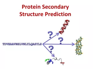
Protein Structure -Primary and Secondary Structure-. Chapter 3 (Page 96-97; 106-107) Chapter 4 (Page 116-125). 1. Protein Structure. Provides deep insight into: P rotein function and stability The evolutionary relatedness of all proteins, and more fundamentally of all organisms Disease

E N D
Protein Structure -Primary and Secondary Structure- Chapter 3 (Page 96-97; 106-107) Chapter 4 (Page 116-125)
1. Protein Structure Provides deep insight into: • Protein function and stability • The evolutionary relatedness of all proteins, and more fundamentally of all organisms • Disease Though it can be quite complex and diverse, protein structure can be broken down to four levels of structure.
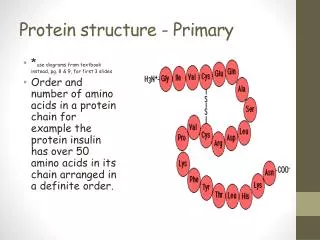
2. Primary Structure In our discussions of amino acids, amide and disulfide bond formation, and protein sequencing we have essentially alluded to the primary structure of proteins. • The sequence of amino acid residues (i.e. HAYP…) • A description of all COVALENT bonds linking amino acids • Bond strength ~200 to 460 kJ/mol for covalent bonds in proteins
2. Primary Structure C. Proteins can be polymorphic • 20-30% of proteins in humans have amino acid sequence variants • The variations can have little or no effect on protein function especially if conservative variations such as Asp substituting for a Glu
2 I. Protein Homology dictates Evolutionary Ties • Protein homology refers to the degree to which the AA sequences of proteins is the same • Typically measured in % homology • % homology, evolutionary relatedness B. AA sequence can vary considerably yet as long as crucial regions are conserved, proteins may exhibit similar function • Crucial regions such as AA that are important for ligand (or substrate) binding • The conserved primary structure of these regions will not result in comparable activity if other levels of structure are widely different
2II. Arrangements of the Atoms in Primary Structure Covalent bonds place important constraints on the conformation of a polypeptide. • 3 D Structure
2II. Arrangements of the Atoms in Primary Structure B. Peptide C-N bond is shorter than normal C-N bonds C. Six atoms of the peptide group lie in a single plane
2II. Arrangements of the Atoms in Primary Structure D. There are two bond angles resulting from rotation at Cα • ϕ(phi) for N-Cα • Ψ(psi) for Cα-C bond • When ϕ= Ψ = 180°; fully extended conformation • Many angles prohibited due to steric interference between atoms in polypeptide backbone and amino acid side chains ϕ= Ψ = 0 PROHIBITED
The Ramachandran Plot This plot depicts the allowed values for ϕand Ψfor sequences of a particular AA. All plots are similar except for glycine.
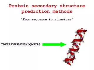
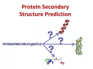
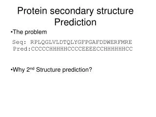
3. Secondary Structure • This level of structure refers to the local conformation of some portion of a protein • Characterized by recurring structural patterns • Linus Pauling and Robert Corey predicted and discovered two of the most prominent secondary structures (1951) even before the first complete protein structure was determined • Demonstrated the importance of NONCOVALENT interactions, particularly hydrogen bonding • Polar C=O and N-H groups of amino acids can engage in H Bonds C=O H-N …
3I. The Alpha (α) Helix • The simplest arrangement the polypeptide chain could assume with its rigid peptide bonds is a helical structure • Stabilized by H bonds between nearby residues • Backbone is tightly wound around an imaginary axis
3I. The Alpha (α) Helix D. The R groups of the AA residues protrude outward from the helix backbone E. Repeating unit • A single turn of the helix, 5.4 Å along the long axis • Includes 3.6 AA residues F. Helical twist is right-handed
3I. The Alpha (α) Helix G. The helix makes optimal use of internal H bonds • 3 to 4 H bonds for each turn • Every peptide bond (except @ N and C-terminus ends) engages in H Bond between H of N atom in amide bond and the O atom of the 4th AA on the amino terminal side H. Predominant secondary structure • Excellent stability • Atoms in close contact Space Filling view
3Ib. Some Amino Acids resist α Helix Structure • Long block of Glu and/or Asp residues (negatively R groups) due to electrostatic repulsion B. Long block of Lys and/or Arg residues (positively charged R groups) also due to electrostatic repulsion
3Ib. Some Amino Acids resist α Helix Structure C. Glyhas more conformational flexibility and adopts a coiled structure different from αHelix D. Pro introduces a destabilizing kink due to its rigid ring structure • No N-Cαrotation
3Ic. Stabilizing Interactions in αHelices Two AA will interact 3 AA units away through: A. Ion pairing between positively charged and negatively charged AA B. Hydrophobic interactions between aromatic AA Ion pairing or Hydrophobic Interactions
3Id. α Helix Net Dipole The polypeptide of an αHelix tends to have a net dipole that is extended through the H bonds. • Amino terminus has partial positive charge • Negatively charged AA at this end • Carboxy terminus has partial negative charge • Positively charged AA at this end
3II. The Beta (β) Sheet • More extended conformation of polypeptide chains • The planarity of the peptide bond and tetrahedral geometry of the -carbon create a pleated sheet-like structure • Side chains protrude from the sheet alternating in up and down direction
3II. The Beta (β) Sheet D. Sheet-like arrangement of backbone is held together by hydrogen bonds between the backbone amides in different strands • Parallel Orientation • Strands have same amino-to-carboxyl orientations • Bent H-bonds • 6.5 Å repeat units
3II. The Beta (β) Sheet • Antiparallel Orientation • Strands have opposite amino-to-carboxyl orientations • Straight H-bonds (Stronger) • 7.0 Å repeat units
3II. The Beta (β) Sheet E. When two or more chains are layered together, the R groups of the AA must be relatively small • Proteins like β-Keratins have very high Gly and Ala content F. The individual chains can be nearby or far in the protein
3III. The Beta Turns • turns occur frequently whenever strands in sheets change the direction • The 180° turn is accomplished over four amino acids • The turn is stabilized by a hydrogen bond from a carbonyl oxygen to amide proton three residues down the sequence • Prolinein position 2 or glycine in position 3 are common in turns
3IIIb. Two types of Beta Turns • Type 1 occurs twice as much as Type 2 • Proline occurs at residue 2 because it can assume the cis configuration (6% frequency) • B. Type 2 always has a Gly as the 3rd residue
4. Spectroscopic Detection of Secondary Structure Because proteins are made of amino acids, which are optically active, they exhibit circular dichroism (CD) spectroscopy • CD measures the molar absorption difference of left- and right-circularly polarized light: = L– R • Characteristic signals are produced based on the environment of the chiral molecule • CD signals from peptide bonds depend on the chain conformation
4. Spectroscopic Detection of Secondary Structure D. CD in the ultraviolet region (UV), 190 to 250 nm, is used to investigate the secondary structure of proteins E. % of α Helix and β Sheet can be determined from the CD spectrum • α Helix, CD spectrum will appear more αHelix in form F. Change in CD spectrum indicates a structural change • Typically from one structure to a random coil