Download
1 / 57
570 likes | 588 Views
Explore how a few atoms can control macroscopic biological functions through the structure and function of ion channels. Learn about the current-voltage relationship in biological systems and the critical role of ion channels in cellular activities. Discover the challenges in modeling and engineering ion channels for therapeutic interventions.

E N D
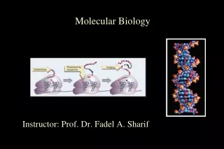
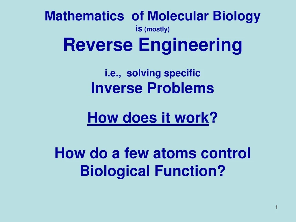
Mathematics of Molecular Biology is (mostly) Reverse Engineering i.e., solving specific Inverse Problems How does it work? How do a few atoms control Biological Function?
Ompf G119D A few atoms make a BIG Difference Glycine replaed by Aspartate Structure determined by Raimund Dutzler in Tilman Schirmer’s lab Current Voltage relation by John Tang in Bob Eisenberg’s Lab
Mathematics of Molecular Biology How does it work? How do a few atoms control (macroscopic) Biological Function? Inherently multiscale Inherently nonequilibrium
How do a few atoms control (macroscopic) Biological Function? Inherently multiscale. Inherently nonequilibrium. Inherently involves macroscopic boundary conditions. Inherently involves nonideal ionic solutions.
+ ~30 Å Ion Channelsare theValves of CellsIon Channels are the Main Controllers of Biological Function Ions in Water* are the Selectivity Different Ions carry Different Signals Liquid of Life *Pure H2O is toxic to cells & proteins Na+ Hard Spheres Ca++ Chemical Bonds are lines Surface is Electrical Potential Redis negative (acid) Blueis positive (basic) K+ 3 Å 0.7 nm = Channel Diameter Figure of ompF porin by Raimund Dutzler
Valves Control Flow Classical Theory: NOT designed for flow Thermodynamics, Statistical Mechanics do not allow flow Rate Models are inconsistent with Maxwell’s Eqn (Kirchoff Law) (if rate constants are independent of potential)
Tutorial What is the biological data?
The Membrane The Cell
Closed Channel Open Channel ION CHANNELS – Biological Role Ion channels coordinate contraction of cardiac muscle making the heart a pumpIon channels coordinate contraction in skeletal muscleIon channels control all electrical activity and produce nerve signalsIon channels are involved in secretion and absorption in all cells:kidney, intestine, liver, adrenal glands, etc.Ion channels are involved in thousands of diseases and many drugs act on channels Ion channels are proteins with genes (blueprints) manipulated by molecular genetics Ion channels have structures shown by x-ray crystallography in favorable cases
OmpF Biochemist’s View Structure All Atoms View Chemical Bonds are lines Surface is Electrical Potential Red is positive Blue is negative Bob Eisenberg: beisenbe@rush.edu
Single Channel Current open closed Slide from Mike Fill Thanks! Function of SINGLE isolated RyR Channels in Artificial Planar Lipid Bilayers AxoPatchPatch-Clamp Amplifier Designed at Rush Planar Bilayer Ca Fused Vesicle Experimental Chamber Teflon Septa 80-100 µM Diameter
Channel Structure Does Not Change once the channel is open Amplitude vs. Duration Current vs. time Open Closed Open Amplitude, pA 5 pA 100 ms Open Duration /ms Lowpass Filter = 1 kHz Sample Rate = 20 kHz Typical Raw Single Channel Records Ca2+ Release Channel of Inositol Trisphosphate Receptor: slide and data from Josefina Ramos-Franco. Thanks!
Single Channel Currents have little variance John TangRush Medical Center
Goal: Understand Selectivity well enough to Make a Calcium Channel using techniques of molecular genetics, site-directed Mutagenesis
Channels are Selective because Diameter Matters Ions are NOT Ideal Potassium K+ = Na+ Sodium / K+ Na+ 3 Å Ideal Ions are Identical if they have the same charge
+ ~30 Å Channels are SelectiveDifferent Ions Carry Different Signals through Different Channels ompF porin Ca++ Na+ K+ 0.7 nm = Channel Diameter 3 Å Diameter mattersDiameter is the Only Difference between K+ and Na+ In ideal solutions K+ = Na+ Flow time scale is 0.1 msec to 1 min Figure of ompF porin by Raimund Dutzler
Experiments have builtTwo Synthetic Calcium Channels MUTANT ─ Compound Calcium selective Unselective Wild Type As density of permanent charge increases, channel becomes calcium selectiveErev ECa in0.1M1.0 M CaCl2 built by Henk Miedema, Wim Meijberg of BioMade Corp.,Groningen, Netherlands Miedema et al, Biophys J 87: 3137–3147 (2004) Mutants of ompF Porin Designed by Theory Glutathione derivatives Atomic Scale || Macro Scale
Channels are only Holes Why can’t we understand and build them? Where to start? Why not compute all the atoms?
Multiscale Issues Journal of Physical Chemistry C (2010 )114:20719 Three Dimensional (106)3 Biological Scales Occur Together so must be Computed Together This may be impossible in simulations Physicists and Engineers rarely try
Why can’t we understand and build channels? Uncalibrated Simulations will not make devices that actually work Calibration is Hard Work particularly for Non-Ideal systems with Correlations, Finite Size effects, and Flows
Where do we start? Physics ‘As Usual’‘Guess’, Calculate andCheck Crowded Charges
Active Sites of Proteins are Very Charged 7 charges ~ 20M net charge = 1.2×1022 cm-3 liquidWater is 55 Msolid NaCl is 37 M + + + + + - - - - Selectivity Filters and Gates of Ion Channels are Active Sites Physical basis of function OmpF Porin Hard Spheres Na+ Ions are Crowded K+ Ca2+ Na+ Induced Fit of Side Chains K+ 4 Å Figure adapted from Tilman Schirmer
Charge Density22 M EC#: Enzyme Commission Number based on chemical reaction catalyzed #AA: Number of residues in the functional pocket MS_A^3: Molecular Surface Area of the Functional Pocket (Units Angstrom^3) CD_MS+: Charge Density (positive) CD_MS-: Charge Density (negative) CD_MSt: Total Charge density Jimenez-Morales, Liang, Eisenberg
Working Hypothesis Biological Adaptation is Crowded Ions and Side Chains Everything interacts
Working Hypothesis Interactions in Channels come mostly from Finite Size Effects Chemically Specific Propertiescome from Diameter and Charge learned from Doug Henderson, J.-P. Hansen, Stuart Rice, among others…Thanks!
Bulk Solutions: Interactions come mostly from Finite Size Effects Chemically Specific Properties of ions (e.g. activity = free energy per mole) are known to come from interactions of their Diameter and Charge and dielectric ‘constant’ of ionic solution Atomic Detail ‘All Spheres’ Model’= Primitive Implicit Solvent Modellearned from Doug Henderson, J.-P. Hansen, Stuart Rice, among others…Thanks!
Na+ Three Channel Types RyR, CaV= EEEE, andNav= DEKA analyzed successfully* in a wide range of solutions by the ‘All Spheres’ Primitive Model Implicit solvent model of open channel ½ ½ ½ ½ ½ ½ ½ Na+ Na+ Na+ ionsandproteinside chains are hard spheres in this model * Many methods have been used in more than 30 papers since Nonner and Eisenberg, 1998 ½
Solved with Many Methods with similar results Metropolis Monte Carlo MSA(mean spherical approximation SPM (primitive solvent model) DFT (density functional theory of fluids), MC-loc(MC with localized side chains) Non-equilibrium Multiscale DFT-PNP (Poisson Nernst Planck) EnVarA (Energy Variational Approach) DMC Dynamic Monte Carlo NP-LEMC (Nernst Planck Local Equilibrium Monte Carlo) Steric PNP Fermi-Poisson (fourth order PDE); etc.
Best Evidence is from the RyRReceptor Dirk GillespieDirk_Gillespie@rush.edu Gerhard Meissner, Le Xu, et al, not Bob Eisenberg More than 120 combinations of solutions & mutants 7 mutants with significant effects fit successfully
The Geometry • Selectivity Filter • is 10 Å long and 8 Å in diameter • confines four D4899negative amino acids • Four E4900positive amino acids are onlumenal side,overlapping D4899 • Cytosolic distributedcharge Protein Cytoplasm Lumen Protein D. Gillespie et al., J. Phys. Chem. 109, 15598 (2005).
Ryanodine Receptor Pore Fig 3 The RyR1 conduction pathway from Zalk et al, Nature, 2014, 10.1038/nature13950 “b, Scheme … of all the negatively charged residues in the ionic pathway (red dots) and the [other] negatively charged residues”
1. Gillespie, D., Energetics of divalent selectivity in a calcium channel: the ryanodine receptor case study. Biophys J, 2008. 94(4): p. 1169-1184. 2. Gillespie, D. and D. Boda, Anomalous Mole Fraction Effect in Calcium Channels: A Measure of Preferential Selectivity. Biophys. J., 2008. 95(6): p. 2658-2672. 3. Gillespie, D. and M. Fill, Intracellular Calcium Release Channels Mediate Their Own Countercurrent: Ryanodine Receptor. Biophys. J., 2008. 95(8): p. 3706-3714. 4. Gillespie, D., W. Nonner, and R.S. Eisenberg, Coupling Poisson-Nernst-Planck and Density Functional Theory to Calculate Ion Flux. Journal of Physics (Condensed Matter), 2002. 14: p. 12129-12145. 5. Gillespie, D., W. Nonner, and R.S. Eisenberg, Density functional theory of charged, hard-sphere fluids. Physical Review E, 2003. 68: p. 0313503. 6. Gillespie, D., Valisko, and Boda, Density functional theory of electrical double layer: the RFD functional. Journal of Physics: Condensed Matter, 2005. 17: p. 6609-6626. 7. Gillespie, D., J. Giri, and M. Fill, Reinterpreting the Anomalous Mole Fraction Effect. The ryanodine receptor case study. Biophysical Journal, 2009. 97: p. pp. 2212 - 2221 8. Gillespie, D., L. Xu, Y. Wang, and G. Meissner, (De)constructing the Ryanodine Receptor: modeling ion permeation and selectivity of the calcium release channel. Journal of Physical Chemistry, 2005. 109: p. 15598-15610. 9. Gillespie, D., D. Boda, Y. He, P. Apel, and Z.S. Siwy, Synthetic Nanopores as a Test Case for Ion Channel Theories: The Anomalous Mole Fraction Effect without Single Filing. Biophys. J., 2008. 95(2): p. 609-619. 10. Malasics, A., D. Boda, M. Valisko, D. Henderson, and D. Gillespie, Simulations of calcium channel block by trivalent cations: Gd(3+) competes with permeant ions for the selectivity filter. Biochim Biophys Acta, 2010. 1798(11): p. 2013-2021. 11. Roth, R. and D. Gillespie, Physics of Size Selectivity. Physical Review Letters, 2005. 95: p. 247801. 12. Valisko, M., D. Boda, and D. Gillespie, Selective Adsorption of Ions with Different Diameter and Valence at Highly Charged Interfaces. Journal of Physical Chemistry C, 2007. 111: p. 15575-15585. 13. Wang, Y., L. Xu, D. Pasek, D. Gillespie, and G. Meissner, Probing the Role of Negatively Charged Amino Acid Residues in Ion Permeation of Skeletal Muscle Ryanodine Receptor. Biophysical Journal, 2005. 89: p. 256-265. 14. Xu, L., Y. Wang, D. Gillespie, and G. Meissner, Two Rings of Negative Charges in the Cytosolic Vestibule of T Ryanodine Receptor Modulate Ion Fluxes. Biophysical Journal, 2006. 90: p. 443-453.
DFT/PNPvsMonte Carlo Simulations Concentration Profiles Misfit Nonner, Gillespie, Eisenberg Different Methods give Same Results NO adjustable parameters
Error < 0.1 kT/e ChannelPREDICTION 62 measurementsThanks to Le Xu! Note Break in Axis AMFEfor Na+/Cs+ mixturesPredicted before measurements AMFE had not been previously observed Mean ± Standard Error of Mean Bulk Solution 2% error Gillespie, Meissner, Le Xu, et al
Divalents KCl CaCl2 NaCl CaCl2 Misfit CsCl CaCl2 KCl MgCl2 Misfit
KCl Gillespie, Meissner, Le Xu, et al Error < 0.1 kT/e 4 kT/e Misfit
Theory fits Mutation with Zero Charge Theory Fits Mutant in K + Ca Theory Fits Mutant in K Error < 0.1 kT/e 1 kT/e Protein charge densitywild type* 13M Solid Na+Cl- is 37M *some wild type curves not shown, ‘off the graph’ 0M in D4899 1 kT/e Gillespie et alJ Phys Chem 109 15598 (2005)
Calcium Channel More than 35 papers are available at ftp://ftp.rush.edu/users/molebio/Bob_Eisenberg/reprints http://www.phys.rush.edu/RSEisenberg/physioeis.html
Selective Binding CurveL type Ca channel Wolfgang Nonner
‘All Spheres’ Model Side Chains are Spheres Channel is a Cylinder Side Chains are free to move within Cylinder Ions and Side Chains are at free energy minimum i.e., ions and side chains are ‘self organized’, ‘Binding Site” is induced by substrate ions Nonner & Eisenberg
Multiscale Analysis at Equilibrium Solved with Metropolis Monte Carlo MMC Simulates Location of Ionsboth the mean and the variance Produces Equilibrium Distribution of location of Ions and ‘Side Chains’ MMC yields Boltzmann Distribution with correct Energy, Entropy and Free Energy Other methodsgive nearly identical results: Equilibrium MultiscaleMSA (mean spherical approximation SPM (primitive solvent model) DFT (density functional theory of fluids), MC-loc(MC with localized side chains) Non-equilibrium Multiscale DFT-PNP (Poisson Nernst Planck) EnVarA (Energy Variational Approach) DMC Dynamic Monte Carlo NP-LEMC (Nernst Planck Local Equilibrium Monte Carlo) Steric PNP; etc.
Selectivity FilterCrowded with Charge Selectivity Filter O½ Wolfgang Nonner L type Ca Channel + ++ “Side Chains”
Mutation • Na Channel • Ca Channel Same Parameters • E • E • E • A • D • E • K • A Charge -3e Charge -1e 1 0.004 Na+ Ca2+ Na+ Occupancy (number) 0.5 0.002 Ca2+ 0 0 -6 -4 -2 0 0.05 0.1 log (Concentration/M) Concentration/M EEEE has full biological selectivityin similar simulations Boda, et al
Crowded Ions Snap Shots of Contents Radial Crowding is Severe ‘Side Chains’are SpheresFree to move inside channel 6Å Parameters are Fixed in all calculations in all solutions for all mutants Experiments and Calculations done at pH 8 Boda, Nonner, Valisko, Henderson, Eisenberg & Gillespie
Ions in Water are the Liquid of Life They are not ideal solutions Everything Interacts with Everything For Modelers and Mathematicians Tremendous Opportunity for Applied MathematicsChun Liu’s Energetic Variational Principle EnVarA
Calcium Channelhas been examined in ~35 papers, e.g., Most of the papers are available at ftp://ftp.rush.edu/users/molebio/Bob_Eisenberg/Reprints http://www.phys.rush.edu/RSEisenberg/physioeis.html
Selectivity comes from Electrostatic InteractionandSteric Competition for Space Repulsion Location and Strength of Binding Sites Depend on Ionic Concentration and Temperature, etc Rate Constants are Variables
Challenge from leading biophysicists Walter Stühmer and Stefan Heinemann Max Planck Institutes, Göttingen, Leipzig Can a physical theory explain the mutation Calcium Channel into Sodium Channel? DEEA DEKA Sodium Channel Calcium Channel
DEKASodium Channel has very different properties from Ca channel,e.g., ‘binding’ curve,Na+ vs Ca++ selectivity Na+ vs K+ selectivity
Sodium Channel specifically, the Aspartate DAcid Negative Glutamate E Acid Negative Lysine KBasicPositive Alanine A Aliphatic Neutral DEKASodium Channel 6 Å QUALITATIVELY DIFFERENT Properties from the Calcium Channel