Download
1 / 49
650 likes | 2.09k Views
Arterial Blood Gas Analysis. By Mohamed hamdy Assistant lecturer of Anesthesia Ain -Shams Un iversity . Egyptian Resuscitation Council (ERC)Instructor. Why we do Arterial Blood Gas Analysis?. Oxygenation Represented by PaO2 Ventilation Represented by Pa Co2

E N D
Arterial Blood Gas Analysis By Mohamed hamdy Assistant lecturer of Anesthesia Ain-Shams University Egyptian Resuscitation Council (ERC)Instructor
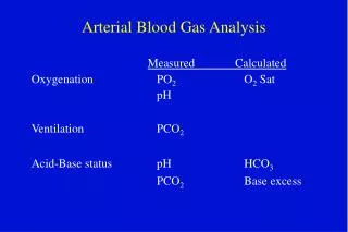
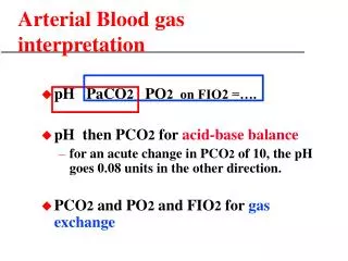
Why we do Arterial Blood Gas Analysis? • Oxygenation • Represented by PaO2 • Ventilation • Represented by Pa Co2 • Acid Base Status • Represented by pH, HCO3 and base deficit. • Hb, Hct, oxygen saturation • Electrolyte e.g. Na+, K+.
Because H+ react highly with cellular proteins resulting in alteration in their function therefore avoiding acidemia and alkalemia by tightly regulation H+ which is essential for normal cellular function.
Calculation of Alveolar Gas Equation and A-a Gradient: PAO2 = FiO2×(Bp-pH2O)-PaCO2/R. = 21×(760-47)-40/0.8 = 100 mmHg. A-a Gradient is alvealo-arterial O2 gradient. A-a Gradient = PAO2 -PaO2 It is normally = Age/4+4. It’s Value: concise D.D of hypoxemia. e.g.: normal A-a Gradient Decrease FiO2 Hypoventilation Ventilation perfusion mismatch Rt to Lt shunting increase A-a Gradient Diffusion abnormality
Approach To Hypoxemia N N A-a Do2 pCO2 FIO2 • PaO2 Alv. Hypo. 100% O2 Corrects V/Q Mis. Diffusion No Correction Shunt
1) Arterial/alveolar ratio(a/A) PaO2/PAO2 Normal value for the a/A ratio is 0.8, meaning that 80% of the alveolar oxygen is reaching the arterial system 2) PaO2/ FIO2 ratio Normal ratio is 550 (a person breathing FIO2 of 1.0 at sea level should have a PaO2 of 550 to 600 mmHg) 3) A-a gradient (on 100% oxygen) PAO2 - PaO2 Where PAO2 is calculated by the alveolar air equation previously presented
4) Arterial-alveolar PCO2 Gradient (a-A PCO2) Arterial PCO2 - Alveolar PCO2 Where Alveolar PCO2 is measured by means of end–tidal PCO2 Normal gradient is an alveolar PCO2 2 mmHg less than arterial, Acute increase reflects increase in physiologic dead space
Sample source and collection:- • Arterial blood sample is common utilized clinically but venous blood may be useful in determining acid base status. • Blood sample should be in heparin coated syringe. • The sample should be analyzed as soon as possible. • Air bubble should be eliminated. • The syringe should be capped and placed in ice.
Problem associated with obtaining ABG: Arterial puncture may result in acute hyperventilation. To minimize that: we should use local anesthetic with small needle. When would you withdraw ABG sample after beginning or stopping O2 supplementation? In absence of significant lung disease we should wait from 5-7 minutes before withdraw ABG sample while patient with obstructive lung disease we should wait 25 min.
Interpretation of ABG • Normal blood gas values:
What is PH? PH is –ve log of H+ concentration. Henderson-Hasselbalch EquationpH = pK’a + log ([HCO3] / 0.03 x pCO2) or more simply: The Henderson equation: [H+] = 24 x ( pCO2 / [HCO3] )
STEPS for interpretation of ABG STEP 1: Determine if numbers fit: H+ = H+ = (7.8-PH)×100. • The Rt side of the equation should be within 10% of the Lt Side. If not so: • Another ABG • Chemistry panel for HCO3 should be done.
Determine if: Acidemia (PH<7.37) OR Alkalemia(PH >7.44) is present. STEP 2: STEP 3: Identify primary disturbance: PH Increase Decrease Alkalosis Acidosis Look at PCO2 Increased Decrease Increased Decreased Metabolic Alkalosis Respiratory Alkalosis Respiratory acidosis Metabolic acidosis
STEP 4: Look at the direction of the change of HCO3/PCO2: • If it is in the same direction it is either simple or mixed change. • But if it is in the opposite direction so it is mixed change. STEP 5: Calculate rate of change of Hco3 and co2 (Expected compensation)
Determine the Anion Gap • The Anion Gap [(Na+) + (K+)] – [(Cl-) + (HCO3-)] • The normal A.G is 12meq ± 4. Normal A.G (Hyperchloremic Acidosis) • ↑ GIT loss of HCO3 as in: diarrhea, high output fistula • ↑ renal HCO3 loss as in: RTA(I, II) • TPN. • ↑ CL containing acids. • Wide Anion Gap Acidosis (↑ endogenou non-volatile acid) • Keto Acidosis • Uremia • Lactic Acidosis • Salicylism • Toxins : Methanol,Paraldehyde,Ethylene glycol
All anions and cations Na+ + UC = (Cl-) + (HCO3-) + UA Na+–(Cl-) + (HCO3-) = UA – UC
Calculation of AG in urine: • Urine AG = ( Na+ + K+) – CL- • In a patient with a hyperchloraemic metabolic acidosis: • A negative UAG suggests GIT loss of bicarbonate (egdiarrhoea) • A positive UAG suggests impaired renal distal acidification (ie renal tubular acidosis).
STEP 6: • If there is metabolic acidosis calculate the A.G • Anion Gap = Na+ - (Cl- + HCO3-) = 12meq ± 4. • Corrected A.G = observed A.G + 2.5 (normal albumin - measured albumin). • If the anion gap ↑ proceed to step 7.
STEP 7: If the anion gap metabolic acidosis is present we should evaluate for additional metabolic disorder because the elevation of anion gap above normal ∆ AG = (AG-12) should be buffered by HCO3. Adding ∆AG to current HCO3 will yield the corrected Hco3 which should be normal value 24 meq/l unless there is another disorder present. Corrected HCO3 = current HCO3 (measured) +∆A.G (Normal value 24 meq/l) • If corrected HCO3 >24 → metabolic alkalosis is also present • If corrected HCO3 <24 → a non gap metabolic acidosis is also present • If corrected HCO3 = 24 → it is pure gap metabolic acidosis.
Final step: Be sure that the interpretation of blood gas is consistent and correlated with the clinical picture of the patient.
Practice is not a magic D.R Mohamad Hamdy Resident in Anaesthesiology & ICU department AIN SHAMS University
Case 1 • A 75-year-old man presents to the ED after a witnessed out of hospital VF cardiac arrest. • Arrived after 10 minutes, CPR had not been attempted. • The paramedics had successfully restored spontaneous circulation after 6 shocks. • On arrival the man is comatose with a GCS of 3 and his lungs are being ventilated with 50% oxygen via ET tube. • He has a ST with rate of 120 min-1 and a blood pressure of 150/95 mmHg.
ABG Analysis reveals: • FiO2 0.5 • pH 7.10 • PaCO2 6.0 kPa (45 mmHg) • PaO2 7.5 kPa(56 mmHg) • HCO3- 14 mmol l-1 • BE - 10 mmol l-1
Case 2 • A 65-year-old man with severe COPD has just collapsed in the respiratory high-care unit. • On initial assessment he is found to be apnoeic but has an easily palpable carotid pulse at 90 min-1. • A nurse is ventilating his lungs with a BVM and supplementary O2 (with reservoir)
ABG Analysis reveals: • FiO2 0.85 (estimated) • pH 7.20 • PaCO2 (80 mmHg) • PaO2 (147 mmHg) • HCO3- 40 mmol l-1 • BE + 14 mmol l-1
Case 3 • A 75-year-old lady is admitted to the ED following a VF cardiac arrest, which was witnessed by the paramedics. • A spontaneous circulation was restored after 4 shocks, but the patient remained comatose and apnoeic. • The paramedics intubated her trachea, and on arrival in hospital her lungs are being ventilated with an automatic ventilator using a tidal volume of 900 ml and a rate of 18 breaths min-1.
ABG Analysis reveals: • FiO2 1.0 • pH 7.60 • PaCO2 2.65 kPa (20 mmHg) • PaO2 25.4 kPa (192 mmHg) • HCO3- 20 mmol l-1 • BE -2 mmol l-1
Case 4 • An 18-year-old male insulin dependent diabetic is admitted to the ED. • He has been vomiting for 48 hours and because he was unable to eat, he omitted his insulin. • He has a ST at a rate of 130 min-1 and his blood pressure is 90/65 mmHg. • He is breathing spontaneously with deep breaths at a rate of 35 min-1 and is receiving oxygen 6 l min-1 via O2 mask. His GCS is 12 (E3, M5, V4).
ABG Analysis reveals: • FiO2 0.4 • pH 6.79 • PaCO2 (14 mmHg) • PaO2 (129.2 mmHg) • HCO3- 4.7 mmol l-1 • BE - 29.2 mmol l-1
Case 5 His vital signs are: • Heart rate 120 min-1 – sinus tachycardia – warm peripheries • Blood pressure 70/40 mmHg • Respiratory rate 35 breaths min-1 • SpO2 on oxygen 92% • Urine output 50 ml in the last 6 hours • GCS 13 (E3, M6, V4)
ABG Analysis reveals: • FiO2 0.4 (approx) • pH 7.12 • PaCO2 4.75 kPa (36 mmHg) • PaO2 8.2 kPa (62 mmHg) • HCO3- 12 mmol l-1 • BE - 15 mmol l-1
Case 6 Which patient is more hypoxemic, and why? • Patient A: pH 7.48, PaCO2 34 mm Hg, PaO2 85 mm Hg, SaO2 95%, Hemoglobin 7 gm% • Patient B: pH 7.32, PaCO2 74 mm Hg, PaO2 55 mm Hg, SaO2 85%, Hemoglobin 15 gm%
Patient A: Arterial oxygen content = .95 x 7 x 1.34 = 8.9 ml O2/dl • Patient B: Arterial oxygen content = .85 x 15 x 1.34 = 17.1 ml O2/dl • Patient A, with the higher PaO2 but the lower hemoglobin content, is more hypoxemic.
Case 7 The PO2 in a cup of water open to the atmosphere is always higher than the arterial PO2 in a healthy person (breathing room air) who is holding the cup. True or False
Case 8 A patient is admitted to the ICU with reabeted vomiting the following BLOOD GASES pH: 7.40PCO2: 38HCO3: 24PO2: 72 ELECTROLYTES, BUN & CREATININE Na: 149K: 3.oCl: 100CO2: 24BUN: 110 Creatinine: 8.7 What is(are) the acid-base disorder(s)?
The patient was both uremic (causing metabolic acidosis) and had been vomiting (metabolic alkalosis).
Case 9 55 yrs old pt. who drink fifth of wesky per day has 2 wks history of diarrhea , Anion gapis 20, HCO3 = 10, PH = 7.30, PO2 =0.90 mmHg, PCO2 = 30 mmHg. What is(are) the acid-base disorder(s)?
Case 10 25 yrs pt. come to ER with fever, abd. pain , vomiting, with the history of migrane PH = 7.33, PCO2 = 8mmHg, PO2 = 80 mmHg, HCO3 = 4, Sodium = 140 mmol, K = 3 mmol, CL = 108 mmol. What is(are) the acid-base disorder(s)?
“Life is a struggle, not against pharama, not against the physics, not against Anesthesia Exame, but against hydrogen ions.“ M. Hamdy