Download
1 / 117
1.17k likes | 1.33k Views
Host Defenses. Microbiology 2314. A Healthy Host Has a Variety of Defenses to Prevent Infection. Definitions. Resistance - Ability to ward off disease. Susceptibility - Lack of resistance. Two Types of Resistance. Nonspecific Resistance / Defense Against All Invaders.

E N D
Host Defenses Microbiology 2314
A Healthy Host Has a Variety of Defenses to Prevent Infection
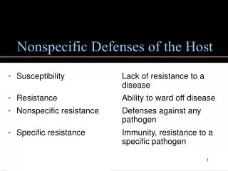
Definitions • Resistance - Ability to ward off disease. • Susceptibility - Lack of resistance.
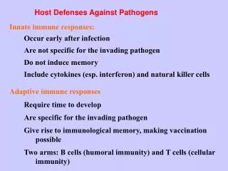
Nonspecific Resistance / Defense Against All Invaders • First Line of Defense 1. Skin 2. Mucus Membranes • Second Line of Defense 1. Phagocytes 2. Inflammation 3. Fever
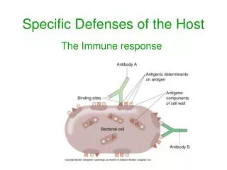
Specific Resistance / Defense Against Specific Microorganisms • Antibodies • Lymphocytes (B and T Cells)
Skin Structure and Composition (Waterproof Keratin) Provide Resistance to Infection. It is not a great place for bacteria to live… Dries Sheds Secretes
Epidermis of the Skin • Epidermis consists of four layers. • Top layer is dead • 15-40 Rows of Dying Cells • Keratin a waterproof protein • pH of 3-5 Symbiotic bacteria living on the skin decompose dead skin cells. The process results in a strong odor as the number of bacteria increase.
Fungus Can Penetrate Keratin to Cause Infection When Excessive Moisture is Present
Best Areas for Organisms to Occupy are Scalp Ears Underarms Genital Regions Why?
Lachrymal Apparatus Protects the eyes from irritating substances and microorganisms. The conjunctiva has only a small number of bacteria present due to continuous blinking and lachrymal secretions which contain bactericidal substances.
The lacrimal glands underneath the skin of the upper eyelids make a fluid that is mostly salt and water. This salty water gets to the eye through small openings inside the upper eyelids.
When the eyelid blinks, the watery liquid is spread across the eye. There are other glands on edges of the eyelids that make oils. The most important of these glands are called the meibomian glands. The oils from these glands actually float on top of the watery fluid in the tears. This keeps the water from evaporating too quickly.
Some of the oils stay along the edge of the eyelid, and they help keep the tears from "leaking" over the eyelashes. If there is not enough of these oils, tears will keep overflowing from the eyes. Oddly enough, a problem with the meibomian glands can lead to overflowing tears and dry eye syndrome at the same time
Salivary Glands Produce Saliva Saliva Washes Microorganisms from Teeth and Gums
Saliva isn’t always enough. S. mutans Secretes Sticky Polysaccharide Plaque
Tooth decay (dental caries) was not a major problem before the fateful year of 1886. Do you remember why 1886?
That was the year that Coca Cola was first invented and marketed.
European Teeth Teeth in skulls from Europeans prior to the 1500’s showed remarkably well-reserved teeth. Once sugar was introduced into the European diet, teeth deteriorated quickly and tooth decay became a widespread disease.
Defensive Body Secretions • Tears • Saliva • Mucus • Vaginal Secretions • Nasal Secretions • Sebum • Perspiration
Mucus traps many microorganisms that enter the respiratory and gastrointestinal tracts. Snotty noses are a result of the body trying to rid the system of the trapped microorganisms.
The nostrils are heavy with bacteria but the sinuses and lungs are usually sterile.
The flow of urine moves microorganisms out of the urinary tract. The kidney and bladder are usually sterile.
What is Sebum? Sebum is an oily/fatty substance secreted from the sebaceous glands (unsaturated fatty acids) that inhibits the growth of pathogenic bacteria. Constituent % by weight Glycerides and free fatty acids 57.5 Wax esters 26.0 Squalene 12.0 Cholesteryl esters 3.0 Cholesterol 1.5
Unfortunately, Some Bacteria Have Adapted and Now Metabolize Sebum
Perspiration • Washes Microorganisms Off Skin • Maintains Body Temperature • Eliminates Cellular Wastes • Contains Lysozyme • Lysozyme is Most Effective Against Gram Positive Bacteria
Increased Perspiration Does Increase the Growth of Some Bacteria.
Lysozyme • Found In 1. Tears 2. Salava 3. Nasal Secretions 4. Perspiration
The Acidity of Gastric Juice Prevents Most Microbial Growth in the Stomach • Mucus, HCl, Enzymes • pH 1.2 - 3.0 • No effect on Clostridium botulinum • No effect on Staphylococcus aureus
Ulcers • 4 Million Americans Have Ulcers Annually • H. pylori is Found in Almost 50% of the Population • That 50% Could Either Have Gastritis or Ulcers
Why is H. pylori able to survive in the acidic environment of the stomach if it isn’t an acidophil?
PhagocytosisThe Ingestion of Microorganisms or Particulate Matter by a Cell We frequently see this when WBC’s engulf bacteria.
Phagocytosis • Attraction (Chemotaxis) • Attachment (Opsonization / Coating with Protein) • Ingestion • Digestion (Lysosomal Enzymes and Oxidizing Agents) • Expulsion
The Mechanism of Phagocytosis Chemotaxis is the process by which phagocytes are attracted to microorganisms.
Pus is the accumulation of damaged tissue and dead microbes and white blood cells.
An accumulation of pus in the front of the eye. In this image, the pus is seen as a pool of whitish fluid between the iris and cornea.
White Blood Cells (Leukocytes) Macrophages are Mature Monocytes that are Phagocytic in Nature Histiocytes are a special type of macrophage fixed in a particular tissue 1. Kupffer Cells / Liver 2. Alveolar macrophages / Lungs
WBC - Neutrophils • Increased / Bacterial Infection • Normally 50-70 • Two Types 1. Segmented (Mature) 2. Banded (Less Mature)
WBC - Basophils • Allergic Reactions • Leukemias • Normally 0-1
WBC - Eosinophils • Worm Infections • Skin Disorders • Scarlet Fever • Normally 1-5
WBC - Lymphocytes • Viral Infections • German Measles • Whooping Cough • Syphilis • Normally 20-30
WBC - Monocytes • Recovery from Infections • Fungal, Rickettsial, Protozoal, Infections • Normally 2-6
Differential WBC Count (100 Cells) • Neutrophils 50-70 • Lymphocytes 20-30 • Monocytes 2 - 6 • Eosinophils 1 - 5 • Basophils 0 - 1 Wright's stain is a technique that is used to make the differences between cells visible under light microscopy. It is used in the examination of peripheral blood smears and bone marrow aspirates.