Download
1 / 216
2.16k likes | 2.53k Views
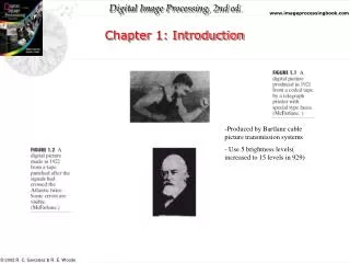
Chapter 1. introduction. 1.1. Definition A. Biochemistry: Biological Chemistry, Physiological Chemistry Chemistry General Chemistry Organic Chemistry—Last parts Inorganic Chemistry Physical Chemistry Quantum Chemistry B. Biochemistry

E N D
Chapter 1. introduction 1.1. Definition A. Biochemistry: Biological Chemistry, Physiological Chemistry Chemistry General Chemistry Organic Chemistry—Last parts Inorganic Chemistry Physical Chemistry Quantum Chemistry B. Biochemistry Structures of Biomolecules Bioenergetics Functions of Biomolecutes Biomolecules: Large and small molecules Large ones: Macromolecules like DNA, RNA , proteins and polysaccharides Bioenergetics: Energy flow in the living cells C. Conformation: 2 conformations of ethane Eclipsed Staggered
A. Starch and Cellulose: Homopolymers of Glucose B. Proteins: Heteropolymers of Amino Acids : Acids containing amine groups C. Nucleic Acids: Heteropolymers of Nucleotides( dAMP AMP, dGMP GMP, dTMP UMP, dCMP, CMP) 1.4. Organelles, Cells and Organisms A. Archaebacteria: Methane bacteria B. Prokaryotes: Pro-Before, Karyon- nuts or kernel Page 16 Table 1.1
C. Eukaryotes: Eu- Good or well Human body digestive system Liver Hepatocytes Nucleus Chromatin DNA Nucleotides Base, sugar and phosphate C, H, O, N Page 19 Fig. 1.11
1.5 Life at the Extremes: Page 16 Window on Biochemistry 1.6 Handling cell components: Page 24 Window
Chapter 2. The flow of biological information: Cell communication 2.1. Brief Image of information flow: Page 30 Fig. 2.1 Transcription Translation
2.2. Storage of Biological information in DNA Genome: The total genetic informational content for each cell Exact duplication Expression of stored information DNA molecules Watson and Crick in 1952: Double helix structure Complementary base pairs by specific hydrogen bondings: C-G and A-T C-G: triple hydrogen bonds A-T: double hydrogen bonds Bases: inside the helix , the backbone : sugar and phosphate Human Genome Project To map and sequence the estimated 3 billion nucleotide base pairs Other living organisams: Bacillus subtilis, Caenorhabditis elegans, Yeast, Arabdopsis thaliana, Rice 30,000-40,000 genes in the human genome Proteomics: The name given to the broad field investigating the thousands of protein products from the genome Bioinformatics: Computer applications to organize the mass of nucleic acid sequence data and studying relationships between protein sequence and structure.
2.3 Replication • 5’ to 3’, Semi conservative replication • DNA polymerases • Polymerase Chain Reaction (PCR) • 2.4 Transcription • Double helix DNA, RNA polymerase • rRNA, mRNA, tRNA: Page 35 Table 2.1
2.5 Translation • Genetic code: triplet code Page 38 Fig. 2.7
Exons and introns Introns are absent in prokaryotes RNA processing: Page 38 Fig. 2.8 Catalytic RNA
2.6 Errors in DNA Processing • DNA mutations: sickle cell anemia and other inborn metabolic errors • 2.7 Information flow through cell membranes • A. Signal transduction: • B. Second Messengers: cAMP, cGMP, Ca2+ • 2.8 Drug design: • Page 44 Fig 2.11 • A. Interference of Protein synthesis including transcription and translation • B. Receptor binding to block the initiation • C. Disruption of Signal pathway
Chapter 3 Biomolecules in Water Water content: Page 48 Table 3.1, 71-83%
3.1 Water the Biological Solvent Weaker interactions: noncovalent bonds- 1-30kj/mole (cf. 350kj/mole for C-C bond) Page 50 Table 3.2 Reversible and specific Van der Waals forces Ionic bonds Hydrogen bonds Hydrophobic interactions
B. The structure of water Page 51 Fig. 3.1, sp3 orbitals
3.2 Hydrogen bonding and Solubility • Physical properties of water: Page 53 Table 3.3 , Fig. 3.5 Solvation: Page 54 Fig. 3.6 Amphiphilic Hydrophobic interaction Micelles
3.3. Cellular Reactions of Water A. Ionization of water
B. pH and pK pH = -log [H+] , pKa = -log Ka pH values of some natural fluids: Page 58 Fig 3.10 HA H+ A- Ka = [H+][A-]/[HA] : Acid Conjugate Base pH = pKa log [A-]/[HA]: Handerson-Haselbalch Equation H3PO4 H+ H2PO4-1 pKa1=2.14 H2PO4-1 H+ HPO4-2 pKa2 = 7.20 HPO4-2 H+ HPO4-3 pKa3 = 12,4 Titration curve CH3COOH + NaOH = CH3COONa + H2O Acid Conjugate base pH = pKa log [A-]/[HA] If [A-]/[HA]= 1 , log [A-]/[HA] = 0. So pH = pKa 3.4 Buffer system Acid –base conjugate pairs Buffering blood and other cellular fluids: Page 62, Window, Table 3.5 Buffer exercises : Acetate buffer, Phosphate buffer
Chapter 4. Amino acids, Peptides and Proteins: Proteins architecture and Biological Functions • 4.1. The amino acids in proteins • D- and L-form: Mirror image ( Enantiomers) • Classification and Properties
C. Modified amino acids: 4-hydroxy proline, 5-hydroxy lysine D. Reactivity of amino acids Reagents reacting with amine group: Page 77 Fig 4.9
4.2. Polypeptides and Proteins Peptide bonds: Page 101 Fig 5.4 Average molecular weight of all amino acids in the polypeptides: 110
B. Amino terminus ( N-terminus), Carboxyl terminus( C-terminus) C. Peptidases, or Proteases D. Oligopeptides: Glutathione(3AA), Enkephalin (5AA), oxytocin( 9AA), vasopressin (9AA), insulin (51 AA) E. Classification Shape- globulins, fibrous proteins Function- Page 79-82 Components- Simple proteins, Complex( Conjugated) proteins ( glyco-, lipo- , metalo- chromo-, phospho-, nucleo-) 4.3 Four levels of protein structure A. Primary structure: Amino acid sequence and disulfide bonds B. Secondary Structure: Conformation of neighboring amino acids: -helix,: Right handed or left handed coil: One turn of the helix: 0.54 nm and 3.6 residues: Page 102 Fig. 5.5 -pleated sheet: parallel, antiparallel: Silk Fibroin or proteins of spider webs Bends and loops: proline and glycin at the bend: Extended bends: loops Super secondary structure or Motifs ( Domains) The individual elements of secondary structures are often combined into stable, geometrical arrangements. , ,,
C. Tertiary structure: Conformation of distant amino acids Page 100 Fig. 5.3
Primary structure determines the tertiary structure. Denaturing, Renaturing, Native proteins Page 109 Fig 5.14 D. Quaternary structure Monomeric, oligomeric Subunits
4.4. Hemoglobin A. Heme – globin B. Heme binding to globin C. Oxygen binding: Page 115, Fig. 5.19 Sigmoidal curve—Positive cooperation( Myoglobin: Hyperbolic) D. Bohr effect: Page 115 Fig. 5.20 H+ and CO2 decrease the affinity of hemoglobin for oxygen molecule 4.5. Fashionable hair: Page 114, Windo
Chapter 5. 6. Enzymes 1 • 6.1 Biological catalysts • A. Catalysts: Page 127 Fig 6.2
B. Cofactor- in addition to the protein component, another chemical entity , in order to function properly. Metal ions- Zn+2, Mg+2 Coenzymes: Orgnometallic molecules C. Prosthetic groups: cofactor covalently bonded to the proteins Holoenzyme: protein + cofactor Apoenzyme: without cofactor E. Classification : Page 129 Table 6.2
6.2 Enzyme Kinetics • A. Michaelis Menten equation derivatisation • k2 k4 • E + S ES E + P • k1 k3 • ES formation rate: k1[E][S] + k4[E][P], often k4 is very small to be neglected. • ES degradation rate: k3[ES] + k2[ES] • At Steady state: Rate of formation = rate of degradation • k1[E][S] = k3[ES] + k2[ES] • [E]total = [E] + [ES] • k1([E]total-[ES]) [S] = [ES](k3 + k2) • k1[E]total[S]-k1[ES][S] = [ES](k3 + k2) • k1[E]total[S]= [ES](k3 + k2) + k1[ES][S] • [ES]( k3 + k2 + k1[S]) = k1[E]total[S] • [ES] = k1[E]total[S] / ( k3 + k2 + k1[S]) • [ES] =[E]total[S] / ( k3 + k2)/k1 +[S]) • (k3 + k2)/k1 = Km • [ES] =[E]total [S] / ( Km +[S]) • dP/dt = Vo = k3[ES] =k3[E]total[S] / ( Km +[S]) • k2[E]total = Vmax • Vo= Vmax [S]/(Km + [S])
B. Km: If V = 1/2 Vmax, Km =[S] • C. Turnover number = Vmax/[E]total • D. Lineweaver-Burk Equation • From Vo= Vmax [S]/(Km + [S]) • 1/Vo = (Km + [S])/Vmax[S] • 1/Vo = Km/Vmax 1/Vmax + 1/Vmax Page 134 Fig. 6.5 • E. Characteristics of enzyme reactions • Enzyme concentration, pH, Temperature
6.3 Substrate binding and enzyme action • A. Active site • Lock and Key model; Page 137 Fig 6.10 • Induced fit model: page 138Fig. 6.11 • Transition state analog model: Page 138 Fig. 6.12
B. General Acid-Base catalysis: An Enzyme donates a proton and accept it in the final step Proton transfer to the carbonyl group Water attack to from a tetrahedral intermediate Acceptance of the proton from the intermediate C. Metal ion catalysis: Alkali metal ( Na+, K+) and transition metals ( Mg+2, Mn+2, Cu+2 Zn+2 Fe+2 Fe+3 Ni+2) Hold a substrate properly oriented by coordinate covalent bonds , Page 140 Fig. 6.14 a Polarize the scissile bond or stabilize a negatively charge intermediate. Fig. 6.14 a Participate in biological oxidation-reduction reactions by reversible electron transfer between metal ions and substrate. Fig. 6.14 a
D. Covalent catalysis A nucleophilic functional group on an enzyme reacts to form a covalent bond with the substrate. Page 140, Step 1 and 2
6.4 Enzyme inhibition • A. Reversible and irreversible inhibitors • DIFP, Pesticides • B. Reversible inhibitors: Page 145 Fig 6.19
Competitive inhibitors: Resemble the structure of normal substrate and binds to the active site of the enzyme E + S ES E + P + I EI Noncompetitive inhibitors: Both inhibitor and substrate can bind simultaneously to the enzyme molecule. E + S ES E + P + + I I EI + S ESI Uncompetitive inhibitors: The inhibitor binds only to the ES complex E + S ES E + P + I ESI
C. Protease inhibitor: page 146 Window • Alzheimer’s disease (AD) • -secretase • Amyloid precursor protein ( APP) A-40 + A-42 • AIDS • HIV protease- Viral growth and development: Phe-Pro, Tyr-Pro
Chapter 7: Enzymes II: Coenzymes, Regulation, Abzymes, and Ribozymes 7.1. Enzyme: Coenzyme partners : Vitamins and Coenzymes: Page 157 Table 7.1 Metals as nutrients: Page 161 Table 7.2 7.2. Allosteric enzymes A. Regulatory enzyme E1 E2 E3 E4 E5 A B C D E P Final product inhibition The beginning substrate in the sequence An intermediate formed in the pathway Some external factor such as a hormone All of the above B. Positive and negative Effectors Effectors: Bomolecules influencing the action of an allosteric enzyme. Allosteric enzymes much larger and more complex than nonallosteric enzymes regulatory sties for binding specific efectors Page 165 Fig. 7.2
C. Models to describe allosteric regulation MWC concerted model: TT RR Reaction products Page 165 Fig. 7.5 Sequential model: TT TR RR Reaction products
7.3. Cellular regulation of enzymes A. Covalent modification of regulatory enzymes Phosphorylation of OH group in serine, threonine or tyrosine: Example: Page 168 Fig. 7.7 Attacheeent of an adenosyl monophosphate to a similar OH group Reduction of cystein disulfide bonds
B. Activation by proteolytic cleavage Zymogen active enzyme + peptide: Page 169 Table 7.3 and Fig 7.8 C. Regulation by isoenzymes Enzymes existing in different molecular forms( Multiple forms) Lactate Dehydrogenase (LDH): M4 in muscle H4 in heart
7.4 Site directed mutagenesis and Abzymes and Ribozymes A. amino acid sequence of known enzymes B. Abzymes or Catalytic antibody: Protein antibodies by using transition state analogs as antigens C. Ribozyme: Catalytic RNA : The catalytically active region – 19 -30 nucleotides