Download
1 / 39
390 likes | 568 Views
Ch.7~ MembraneStructure&Function. Fluid mosaic model. Membrane structure, I. Selective permeability Amphipathic~ hydrophobic & hydrophilic regions 1935 Davson Danielli sandwich model Singer-Nicolson: 1972 fluid mosaic model. Cell membrane must be more than lipids… .

E N D
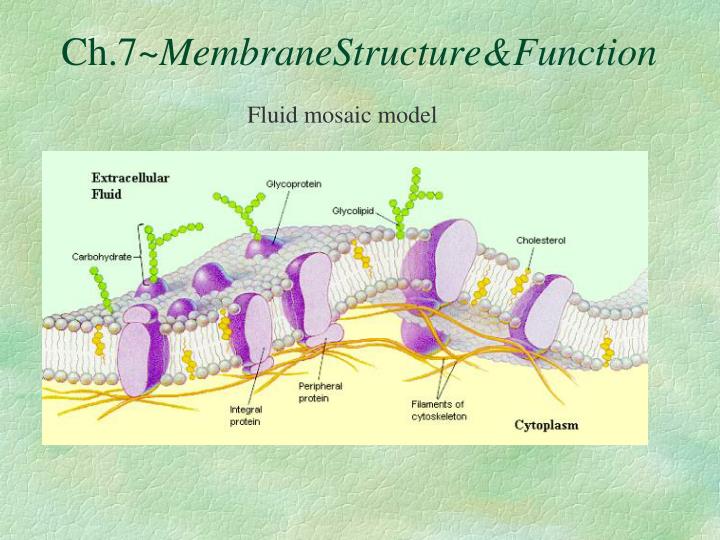
Ch.7~MembraneStructure&Function Fluid mosaic model
Membrane structure, I • Selective permeability • Amphipathic~ hydrophobic & hydrophilic regions • 1935 Davson Danielli sandwich model • Singer-Nicolson: 1972 fluid mosaic model
Cell membrane must be more than lipids… • In 1972, S.J. Singer & G. Nicolson proposed that membrane proteins are inserted into the phospholipid bilayer It’s like a fluid…It’s like a mosaic… It’s the Fluid Mosaic Model!
Glycoprotein Glycolipid Transmembrane proteins Peripheral protein Filaments ofcytoskeleton Membrane is a collage of proteins & other molecules embedded in the fluid matrix of the lipid bilayer Extracellular fluid Phospholipids Cholesterol Cytoplasm 1972, S.J. Singer & G. Nicolson proposed Fluid Mosaic Model
Membrane Structure • A specialized preparation technique, freeze-fracture supports the fluid mosaic model.
Phospholipids Phosphate “attracted to water” • Phosphate head • hydrophilic • Fatty acid tails • hydrophobic • Arranged as a bilayer Fatty acid “repelled by water” Aaaah, one of thosestructure–function examples
Arranged as a Phospholipid bilayer sugar H2O salt • Serves as a cellular barrier / border polar hydrophilic heads nonpolar hydrophobic tails impermeable to polar molecules polar hydrophilic heads lipids waste
Cell membrane defines cell • Cell membrane separates living cell from aqueous environment • thin barrier = 8nm thick • Controls traffic in & out of the cell • allows some substances to cross more easily than others • hydrophobic (nonpolar) vs. hydrophilic (polar)
Permeability to polar molecules? • Membrane becomes semi-permeable via protein channels • specific channels allow specific material across cell membrane inside cell H2O aa sugar salt outside cell NH3
Cell membrane is more than lipids… • Transmembrane proteins embedded in phospholipid bilayer • create semi-permeabe channels lipid bilayer membrane protein channelsin lipid bilyer membrane
Why areproteins the perfect molecule to build structures in the cell membrane?
Proteins domains anchor molecule Polar areas of protein • Within membrane • nonpolar amino acids • hydrophobic • anchors protein into membrane • On outer surfaces of membrane in fluid • polar amino acids • hydrophilic • extend into extracellular fluid & into cytosol Nonpolar areas of protein
Porin monomer H+ Retinal chromophore b-pleated sheets NH2 Bacterial outer membrane Nonpolar (hydrophobic) a-helices in the cell membrane COOH Cytoplasm H+ H+ Examples aquaporin = water channel in bacteria H2O H+ proton pump channel in photosynthetic bacteria function through conformational change = protein changes shape H2O
Many Functions of Membrane Proteins “Channel” Outside Plasma membrane Inside Transporter Enzymeactivity Cell surfacereceptor “Antigen” Cell adhesion Cell surface identity marker Attachment to thecytoskeleton
Membrane Proteins • Proteins determine membrane’s specific functions • cell membrane & organelle membranes each have unique collections of proteins • Classes of membrane proteins: • peripheral proteins • loosely bound to surface of membrane • ex: cell surface identity marker (antigens) • integral proteins • penetrate lipid bilayer, usually across whole membrane • transmembrane protein • ex: transport proteins • channels, permeases (pumps)
Membrane carbohydrates • Play a key role in cell-cell recognition • ability of a cell to distinguish one cell from another • antigens • important in organ & tissue development • basis for rejection of foreign cells by immune system
Simple Diffusion • Move from HIGH to LOW concentration • “passive transport” • no energy needed movement of water diffusion osmosis
HIGH LOW Facilitated Diffusion • Diffusion through protein channels • channels move specific molecules across cell membrane • no energy needed facilitated = with help open channel = fast transport “The Bouncer”
LOW HIGH Active Transport • Cells may need to move molecules against concentration gradient • conformational shape change transports solute from one side of membrane to other • protein “pump” • “costs” energy = ATP conformationalchange ATP “The Doorman”
Active transport • Many models & mechanisms ATP ATP antiport symport
Getting through cell membrane • Passive Transport • Simple diffusion • diffusion of nonpolar, hydrophobic molecules • lipids • HIGH LOW concentration gradient • Facilitated transport • diffusion of polar, hydrophilic molecules • through a protein channel • HIGH LOW concentration gradient • Active transport • diffusion against concentration gradient • LOW HIGH • uses a protein pump • requires ATP ATP
Transport summary simplediffusion facilitateddiffusion ATP activetransport
How about large molecules? • Moving large molecules into & out of cell • through vesicles & vacuoles • Endocytosis~ in • phagocytosis = “cellular eating” • pinocytosis = “cellular drinking” • Recepter mediated • Exocytosis ~out exocytosis
Endocytosis • a cell brings in macromolecules and particulate matter by forming new vesicles from the plasma membrane • 3 types: • 1. phagocytosis, “cellular eating”. • the cell engulfs a particle by extending pseudopodia around it and packaging it in a large vacuole.
Endocytosis • 2. pinocytosis, “cellular drinking”, a cell creates a vesicle around a droplet of extracellular fluid • This is a non-specific process.
Endocytosis • 3.Receptor-mediated endocytosis is very specific in what substances are being transported • triggered when extracellular substances bind to special receptors, ligands, on the membrane surface, especially near coated pits • triggers the formation of a vesicle
The Special Case of WaterMovement of water across the cell membrane
Osmosis is just diffusion of water • Water is very important to life, so we talk about water separately • Diffusion of water from HIGH concentration of water to LOW concentration of water • across a semi-permeable membrane
hypotonic hypertonic Concentration of water • Direction of osmosis is determined by comparing total solute concentrations • Hypertonic - more solute, less water • Hypotonic - less solute, more water • Isotonic - equal solute, equal water water net movement of water
Managing water balance • Cell survival depends on balancing water uptake & loss freshwater balanced saltwater
1 Managing water balance • Hypotonic • a cell in fresh water • high concentration of water around cell • problem: cell gains water, swells & can burst • example: Paramecium • ex: water continually enters Paramecium cell • solution: contractile vacuole • pumps water out of cell • ATP • plant cells • turgid = full • cell wall protects from bursting KABOOM! ATP No problem,here freshwater
Pumping water out • Contractile vacuole in Paramecium ATP
2 Managing water balance I’m shrinking,I’m shrinking! • Hypertonic • a cell in salt water • low concentration of water around cell • problem: cell loses water & can die • example: shellfish • solution: take up water or pump out salt • plant cells • plasmolysis= wilt • can recover I willsurvive! saltwater
3 Managing water balance That’sperfect! • Isotonic • animal cell immersed in mild salt solution • no difference in concentration of water between cell & environment • problem: none • no net movement of water • flows across membrane equally, in both directions • cell in equilibrium • volume of cell is stable • example:blood cells in blood plasma • slightly salty IV solution in hospital I couldbe better… balanced
1991 | 2003 Aquaporins • Water moves rapidly into & out of cells • evidence that there were water channels • protein channels allowing flow of water across cell membrane Peter Agre John Hopkins Roderick MacKinnon Rockefeller
Do you understand Osmosis… .05 M .03 M Cell (compared to beaker) hypertonic or hypotonic Beaker (compared to cell) hypertonic or hypotonic Which way does the water flow? in or out of cell
