Download
1 / 21
210 likes | 635 Views
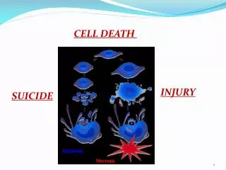
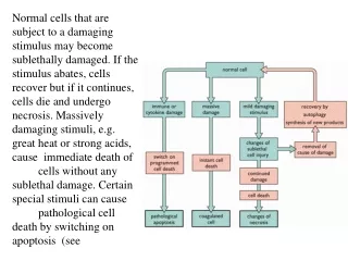
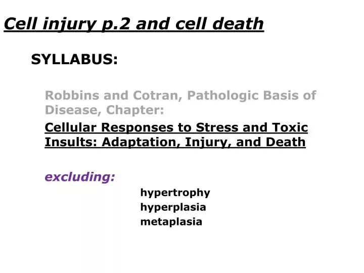
Cell injury p.2 and cell death. SYLLABUS: Robbins and Cotran , Pathologic Basis of Disease , Chapter : Cellular Responses to Stress and Toxic Insults : Adaptation , Injury , and Death excluding : hypertrophy hyperplasia metaplasia. Cell injury p.2 and cell death.

E N D
Cell injury p.2 and cell death • SYLLABUS: • Robbins and Cotran, PathologicBasis of Disease, Chapter: • CellularResponses to Stress and ToxicInsults: Adaptation, Injury, and Death • excluding: • hypertrophy • hyperplasia • metaplasia
Cell injury p.2 and cell death 28Kidney amyloidosis 39Caseous necrosis (tuberculosis, lymph node) 40Liquefactive necrosis (peptic ulcer) 43Balser necrosis (enzymatic fat necrosis) s/35Epidermal cyst 30Leukokeratosis (oral mucosa sample)
Kidney amyloidosis -amyloid deposits in glomeruli and in the walls of blood vessels (metachromatic stain)
Caseous necrosis (tuberculosis, lymph node) • area of necrosis: • amorphous, granular, pink debriswith no visible nuclei or cell contours • elements of TB granuloma: • giant Langhans cells • epithelioid cells • lymphocytes • lymph node structure
Liquefactive necrosis (peptic ulcer) • defect (excavation) caused by the necrosis of the previously pathologically altered tissue • -thin layer of necrotic tissue debris • -underlying inflammatory infiltration
Balser necrosis (enzymatic fat necrosis) -shadowy outlines of necrotic fat cells -basophilic deposits replacing fat in cells -surrounding inflammatory infiltration
Epidermal cyst -normotypical (sometimes flattened) squamous epithelium lining the cyst -keratin masses filling the cyst
Leukokeratosis (oral mucosa sample) -keratinizing squamous epithelium (with granular and keratinized layer) (no skin appendages visible) (.ppt demonstration only, no slides during the microscopy lab)